. 2012 May;30(5):975-87.
doi: 10.1002/stem.1069.

Atmospheric oxygen inhibits growth and differentiation of marrow-derived mouse mesenchymal stem cells via a p53-dependent mechanism: implications for long-term culture expansion

Affiliations

Atmospheric oxygen inhibits growth and differentiation of marrow-derived mouse mesenchymal stem cells via a p53-dependent mechanism: implications for long-term culture expansion

Siddaraju V Boregowda et al. Stem Cells. 2012 May.

Abstract

Large scale expansion of human mesenchymal stem cells (MSCs) is routinely performed for clinical therapy. In contrast, developing protocols for large scale expansion of primary mouse MSCs has been more difficult due to unique aspects of rodent biology. Currently, established methods to isolate mouse MSCs select for rapidly dividing subpopulations that emerge from bone marrow cultures following long-term (months) expansion in atmospheric oxygen. Herein, we demonstrate that exposure to atmospheric oxygen rapidly induced p53, TOP2A, and BCL2-associated X protein (BAX) expression and mitochondrial reactive oxygen species (ROS) generation in primary mouse MSCs resulting in oxidative stress, reduced cell viability, and inhibition of cell proliferation. Alternatively, procurement and culture in 5% oxygen supported more prolific expansion of the CD45(-ve) /CD44(+ve) cell fraction in marrow, produced increased MSC yields following immunodepletion, and supported sustained MSC growth resulting in a 2,300-fold increase in cumulative cell yield by fourth passage. MSCs cultured in 5% oxygen also exhibited enhanced trilineage differentiation. The oxygen-induced stress response was dependent upon p53 since siRNA-mediated knockdown of p53 in wild-type cells or exposure of p53(-/-) MSCs to atmospheric oxygen failed to induce ROS generation, reduce viability, or arrest cell growth. These data indicate that long-term culture expansion of mouse MSCs in atmospheric oxygen selects for clones with absent or impaired p53 function, which allows cells to escape oxygen-induced growth inhibition. In contrast, expansion in 5% oxygen generates large numbers of primary mouse MSCs that retain sensitivity to atmospheric oxygen, and therefore a functional p53 protein, even after long-term expansion in vitro.


Figures

Figure 1
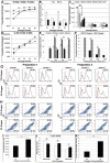
Figure 1. Atmospheric oxygen levels inhibit growth of CD45−ve/CD44+ve bone marrow cells and marrow-derived MSCs purified by immuno-depletion
A–F) Immuno-depleted MSCs plated at the indicated cell densities (500–13,000 cells/cm2) were culture expanded in 21% (A) or 5% (D) oxygen and cumulative cell yields as a function of passage was determined by counting. B) Plotted is the fold increase in MSC number (5000 cell/cm2) as a function of passage for data in (A) and (D). C) MSCs plated at 5000 cells/cm2 were cultured in CCM alone or CCM supplemented with 20 ng/ml FGF2 (FGF2–20), 50 ng/ml FGF2 (FGF2–50), 100ng/ml FGF2 (FGF2–100), 20 ng/ml IGF-1 (IGF-1), or 1000U/ml LIF (LIF) and growth rates (cells/day) in 21% oxygen were quantified as a function of passage by counting. E, F) Population doubling times at P1 and P4 (E) and the total number of population doublings accumulated over four passages (F) were calculated from data in A and D. Doubling times (DT) were determined as DT = (T1–T2)*(Log(2)/Log(Q1/Q2)) where Q1 is the number of cells at T1 and Q2 is the number of cells at T2. Total population doublings at each passage were determined as 2n where n = Log(Q1/Q2)/Log(2). The plotted data (mean ± SD) were determined from experiments run in triplicate. G, H) Two different preparations of whole bone marrow were suspended in CCM supplemented with 10% FBS, expanded in 21% vs. 5% oxygen for 7 days and analyzed by flow cytometry to quantify the percentage of CD45+ve (G, gate illustrated by horizontal line) and CD44+ve/CD45−ve (H, denoted by rectangular gates) subpopulations. Samples were analyzed in triplicate. I) The total yield of plastic adherent cells from bone marrow cultures expanded in 21% vs. 5% oxygen for seven days was determined by counting. J) Plotted are the percentages of CD45+ve, CD45−ve, and CD44+ve/CD45−ve cells in bone marrow cultures quantified by flow cytometry as illustrated in G and H. Note that most CD45+ve cells are also CD44+ve. K). The yield of primary MSCs obtained following immuno-depletion of plastic adherent marrow cultures expanded for 8 days in 21% vs. 5% oxygen was determined by counting. Plotted data (mean ± SD) represent results from at least 5 marrow preparations from each experimental condition. *, p<0.05; **, p<0.01; #, p<0.005; +, p<0.001.
Figure 2
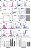
Figure 2. Atmospheric oxygen arrests cell proliferation but does not induce cellular senescence in primary mouse MSCs
A, B) Immuno-depleted MSCs (1000 cells/cm2) were procured and expanded in 5% (A) or 21% (B) oxygen for one passage (P1) and analyzed by flow cytometry for CFSE staining (A) or forward and side scatter (B). C) MSCs cultured as in (A) were sorted based on CSFE label intensity and then analyzed for forward and side scatter. Photomicrographs visually illustrate the distinct morphological differences in populations expanded in 5% vs. 21% oxygen detected by flow cytometric analysis. D) MSCs procured and expanded for four passages (P4) in 5% oxygen were passed an additional time (P5) while maintained in 5% oxygen or switched from 5% to 21% oxygen and then analyzed by flow cytometry for CFSE labeling or forward and side scatter. E) MSCs expanded for four passages (P4) in 21% oxygen were passed an additional time (P5) in 21% oxygen or switched from 21% to 5% oxygen and analyzed as in (D). F, G) Growth rates (F) and population doubling times (G) were calculated for MSCs expanded in 21% oxygen, 5% oxygen, or switch cultures as described above. Plotted values (mean ± SD) represent duplicates from a single experiment. *, p<0.05; **, p<0.01, #, p<0.005.
Figure 3
Figure 3. Exposure to atmospheric oxygen induces p53, TOP2A, and BAX expression and mitochondrial ROS production resulting in oxidative stress and reduced cell viability
Immuno-depleted MSCs were culture expanded in 5% or 21% oxygen for 7 days and biochemical analyses were used to quantify whole cell ROS (A), mitochondrial ROS (B), mitochondrial membrane permeability (C), cell viability (D), protein carbonyl and lipid peroxidase levels (E), and aconitase activity, reduced glutathione and ATP levels (F). All biochemical analyses were performed as described in the experimental procedures and each experiment was conducted using three distinct replicate cultures from each experimental group (5% vs. 21% oxygen cultures). Plotted values (mean ± SD) were determined from four replicates for each sample. D) MSCs were stained with an anti-Annexin V antibody and PI and analyzed by flow cytometry to determine the percentage of early apoptotic, late apoptotic and necrotic cells. G) Cell extracts were prepared from immuno-depleted MSCs that were isolated from bone marrow cultures procured and expanded in either 5% or 21% oxygen for the indicated passage numbers and analyzed by Western blot using an anti-p53 and anti-β-actin antibodies. H) Western blot of the indicated proteins was performed on cell extracts harvested from MSCs expanded in 5% or 21% oxygen at P1 or P2. Western blots were also performed on cell extracts obtained from switch cultures wherein MSCs were expanded for four passages (P4) in 5% oxygen and one additional passage (P5) in 5% oxygen (5/5) or 21% oxygen (5/21). Alternatively, MSCs were expanded to P4 in 21% oxygen and then one additional passage (P5) in 21% oxygen (21/21) or switched to 5% oxygen (21/5). I) MSCs were stained with anti-PCNA and anti-p53 antibodies and analyzed by flow cytometry to determine the percentage of single and double positive cells in populations cultured in 5% or 21% oxygen. *, p<0.05; **, p<0.01, #, p<0.005; +, p<0.001; ++, p<0.0005.
Figure 4
Figure 4. Growth inhibition of MSCs following exposure to atmospheric oxygen is p53 dependent
A, B) Immuno-depleted MSCs (1000 cells/cm2) derived from wild type (A) or p53−/− (B) mice were expanded in 5% or 21% oxygen for one passage (P1) and analyzed by flow cytometry for CFSE staining. C) MSCs from wild type and p53−/− mice were sorted based on CSFE fluorescence and the CSFElow and CSFEHigh fractions were evaluated by flow cytometry based on forward and side scatter. Photomicrographs visually illustrate the distinct morphological differences in populations expanded in 5% vs. 21% oxygen detected by flow cytometric analysis. D) Population doubling times at P1–P5 were calculated for wild type and p53−/− MSCs in 5% or 21% oxygen from data in A. E, F) Cumulative population doublings (E) and cumulative cell number (F) were calculated for from data in D. Doubling times (DT) were determined as DT = (T1–T2)*(Log(2)/Log(Q1/Q2)) where Q1 is the number of cells at T1 and Q2 is the number of cells at T2. Total population doublings at each passage were determined as 2n where n = Log(Q1/Q2)/ Log(2). Plotted values (mean ± SD) represent duplicates from a single experiment. *, p<0.05; **, p<0.01, #, p<0.005.
Figure 5
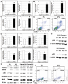
Figure 5. MSCs derived from p53 null mice fail to exhibit increased ROS production and oxidative stress upon exposure to 21% oxygen
Immuno-depleted MSCs from wild type or p53−/− mice were culture expanded in 5% or 21% oxygen for 7 days and biochemical analyses were used to quantify whole cell ROS (A), mitochondrial ROS (B), mitochondrial membrane permeability (C), cell viability (D), protein carbonyl and lipid peroxidase levels (E) and aconitase activity, reduced glutathione and ATP levels (F). All biochemical analyses were performed as described in the experimental procedures and each experiment was conducted using three distinct replicate cultures from each experimental group (5% vs. 21% oxygen cultures). Plotted values (mean ± SD) were determined from four replicates for each sample. D) Immuno-depleted MSCs treated as in A were stained with an anti-Annexin V antibody and PI and analyzed by flow cytometry to determine the percentage of early apoptotic, late apoptotic and necrotic cells. G) Cell extracts were prepared from immuno-depleted MSCs isolated from wild type and p53−/− mice expanded in 5% or 21% oxygen and analyzed by Western blot using anti-p53, anti-MDM2, anti-BAX, and anti-GAPDH antibodies. *, p<0.05; **, p<0.01, #, p<0.005; +, p<0.001; ++, p<0.0005.
Figure 6
Figure 6. Knockdown of p53 ameliorates oxygen-induced changes in ROS production, cell growth, and viability in wild type MSCs
Immuno-depleted MSCs from FVB/N mice were cultured in 5% oxygen and transfected with a p53-specific or scrambled (control siRNA) siRNA. Transfected and control (mock-transfected) cells were then switched to 21% oxygen and 7 days later biochemical analyses were performed to quantify whole cell ROS (A), mitochondrial membrane permeability (B), protein carbonyl (C) and reduced glutathione (D) levels. Each experiment was conducted using three distinct replicate cultures from each experimental group and plotted values (mean ± SD) were determined from four replicates for each sample. E) Cell extracts were prepared from each experimental group at 3 and 7 days post-transfection and analyzed by Western blot using anti-p53, anti-BAX, anti-MDM2 and anti-GAPDH antibodies. F) Cumulative cell number after 7 days expansion of the indicated populations was calculated by counting. G, H) Mock transfected (red line, control) cells and those transfected with a p53-specific (black line) or control siRNA (green line) were labeled with CFSE (G) or Annexin V and PI (H) and examined by flow cytometry. #, p<0.005; +, p<0.001.

Similar articles

Cited by

References

    1. Friedenstein AJ, Chailakhjan RK, Lalykina KS. The development of fibroblast colonies in monolayer cultures of guinea-pig bone marrow and spleen cells. Cell Tissue Kinet. 1970;3:393–403. - PubMed
    1. Digirolamo CM, Stokes D, Colter D, et al. Propagation and senescence of human marrow stromal cells in culture: a simple colony-forming assay identifies samples with the greatest potential to propagate and differentiate. Br J Hematol. 1999;107:275–281. - PubMed
    1. Sotiropoulou PA, Perez SA, Salagianni M, et al. Characterization of the optimal culture conditions for production of human mesenchymal stem cells. Stem Cells. 2006;24:42–47. - PubMed
    1. Schallmoser K, Rohde E, Reinisch A, et al. Rapid large-scale expansion of functional mesenchymal stem cells from unmanipulated bone marrow without animal serum. Tissue Eng Part C Methods. 2008;14:185–196. - PubMed
    1. Iguchi A, Okuyama R, Koguma M, et al. Selective stimulation of granulopoiesis in vitro by established bone marrow stromal cells. Cell Struct Funct. 1997;22:357–364. - PubMed

Publication types

MeSH terms