. 2012 May 15;302(10):L1023-36.
doi: 10.1152/ajplung.00230.2011. Epub 2012 Feb 24.

Inhibiting NF-κB in the developing lung disrupts angiogenesis and alveolarization

Affiliations

Inhibiting NF-κB in the developing lung disrupts angiogenesis and alveolarization

Cristiana Iosef et al. Am J Physiol Lung Cell Mol Physiol. .

Abstract

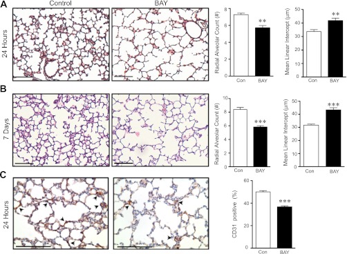
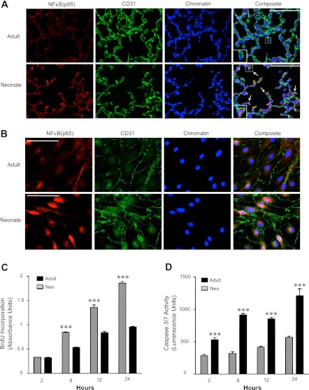
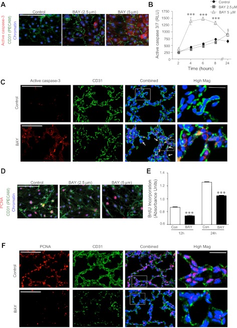
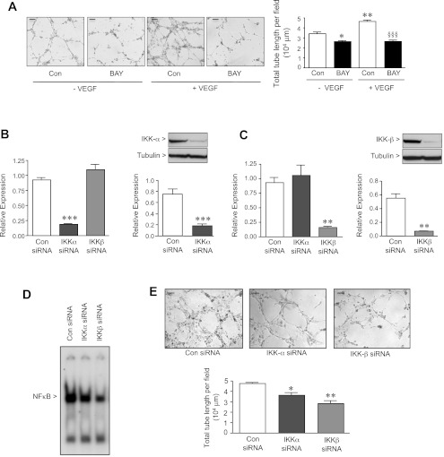
Bronchopulmonary dysplasia (BPD), a chronic lung disease of infancy, is characterized by arrested alveolar development. Pulmonary angiogenesis, mediated by the vascular endothelial growth factor (VEGF) pathway, is essential for alveolarization. However, the transcriptional regulators mediating pulmonary angiogenesis remain unknown. We previously demonstrated that NF-κB, a transcription factor traditionally associated with inflammation, plays a unique protective role in the neonatal lung. Therefore, we hypothesized that constitutive NF-κB activity is essential for postnatal lung development. Blocking NF-κB activity in 6-day-old neonatal mice induced the alveolar simplification similar to that observed in BPD and significantly reduced pulmonary capillary density. Studies to determine the mechanism responsible for this effect identified greater constitutive NF-κB in neonatal lung and in primary pulmonary endothelial cells (PEC) compared with adult. Moreover, inhibiting constitutive NF-κB activity in the neonatal PEC with either pharmacological inhibitors or RNA interference blocked PEC survival, decreased proliferation, and impaired in vitro angiogenesis. Finally, by chromatin immunoprecipitation, NF-κB was found to be a direct regulator of the angiogenic mediator, VEGF-receptor-2, in the neonatal pulmonary vasculature. Taken together, our data identify an entirely novel role for NF-κB in promoting physiological angiogenesis and alveolarization in the developing lung. Our data suggest that disruption of NF-κB signaling may contribute to the pathogenesis of BPD and that enhancement of NF-κB may represent a viable therapeutic strategy to promote lung growth and regeneration in pulmonary diseases marked by impaired angiogenesis.


Figures

Fig. 1.
Fig. 1.
Blocking NF-κB in the neonatal lung disrupts postnatal alveolar development and angiogenesis. A: representative images of hematoxylin and eosin (H&E)-stained lung sections obtained from neonatal mice, 24 h after administration of vehicle or the NF-κB inhibitor, BAY, with morphometric analysis to determine the radial alveolar count (**P = 0.003) and mean linear intercept (**P = 0.008). Data are expressed as means ± SE. B: representative images of H&E-stained lung sections obtained from neonatal mice, 7 days after administration of vehicle or the NF-κB inhibitor, BAY, with morphometric analysis to determine the radial alveolar count (***P = 0.0004) and mean linear intercept (***P = 0.0008). Data are expressed as means ± SE. C: representative images of lung sections, 24 h after administration of vehicle or BAY, immunostained to detect the endothelial-specific marker, CD31 (brown). Quantification of vascular density using a point-counting method expressed as the percentage of CD-31-positive points divided by total parenchymal points, with data expressed as means ± SE and ***P < 0.0001 (n = 5–8 mice per group). Scale bar for all images = 100 μm.
Fig. 2.
Fig. 2.
NF-κB is constitutively active in the developing pulmonary vascular endothelium. A: representative confocal immunofluorescent images from frozen sections of adult and neonatal lung tissue immunostained to detect the NF-κB subunit, p65 (red), and CD31 (green). The presence of nuclear (active NF-κB) is visualized by a pink signal. In the composite panels a single endothelial cell (EC) is magnified and shown in the insets. Arrows indicate examples of CD31-positive cells with p65 in the nucleus. Scale bar = 100 μm. B: representative confocal fluorescent images of adult (top) and neonatal (bottom) pulmonary endothelial cells (PEC) demonstrating localization of the NF-κB subunit, p65 (red) in CD31-labeled (green) ECs. Few p65-positive nuclei were observed in adult PEC, compared with multiple positive nuclei in the neonatal PEC. Scale bar = 50 μm. C: in vitro bromodeoxyuridine (BrdU) incorporation assays performed in adult (solid bars) and neonatal (shaded bars) PEC, 2–24 h after stimulation with endothelial growth media (EGM) containing 5% FBS and EC growth factors (n = 8, with ***P < 0.0001 vs. adult values at that same time point). D: in vitro luminescent caspase-3/7 activity assays performed in neonatal and adult PEC, 2–24 h after serum withdrawal (n = 6, with ***P < 0.001 vs. adult PEC at that same time point).
Fig. 3.
Fig. 3.
BAY treatment effectively decreases active NF-κB in vitro and in vivo. A: representative composite immunofluorescent images from frozen section of lungs obtained from vehicle- and BAY-treated neonatal mice at 24 h, to detect p65 (red), CD31 (green), or chromatin (blue). Scale bar = 100 μm. B: representative composite confocal fluorescent images demonstrating p65 localization (red) in CD31-labeled (green) neonatal PEC 8 h after exposure to either DMSO or BAY (2.5 or 5 μM). Scale bar = 100 μm. C: EMSA using radiolabeled oligonucleotides containing the NF-κB consensus sequence and nuclear extracts obtained from neonatal PEC 4 h after incubation with either vehicle control (DMSO) or BAY (2.5 or 5 μM). D: EMSA with supershift analysis using antibodies against the 5 NF-κB family members and nuclear extracts from neonatal PEC under control conditions, incubated with radiolabeled κB oligo nucleotides (lane 1) Specificity of the band was confirmed by the disappearance of the band with the addition of 100-fold excess of cold oligonucleotide (lane 2). Supershift analysis of the NF-κB complex present in control PEC was then performed (lanes 3–7), with the shift produced by the addition of p65 antibodies (lane 3) labeled as S1, and that produced by p50 antibodies (lane 4) labeled S2. Lanes 5–7 contained supershift reactions using antibodies against p52, cRel, and RelB, respectively.
Fig. 4.
Fig. 4.
Blocking NF-κB increases apoptosis and decreases proliferation in neonatal PEC in vitro and in vivo. A: representative composite fluorescent images of active caspase-3 staining (red) in CD31-labeled (green) neonatal PEC, 8 h after exposure to vehicle (DMSO) or BAY (2.5 or 5 μM). Scale bar = 100 μm. B: in vitro luminescent apoptosis assay to quantify active caspase-3/7 in relative luminescent units (RLU), 2 to 24 h after incubation with vehicle control (DMSO) or BAY (2.5 or 5 μM). Data are means ± SE with n = 4–5 per group. ***P < 0.001 for 5 μM vs. control at 4, 6, and 8 h and vs. 5 μM at 2 h, and §P < 0.05 for 2.5 μM at 24 h vs. 2 h. C: representative confocal fluorescent images from formalin-fixed lung tissue obtained from vehicle- and BAY-treated neonates after 8 h and immunostained to detect active caspase-3 (red), and CD31 (green). Arrows indicate examples of CD31-positive cells with active caspase-3 in the cytoplasm. Scale bar = 100 μm. Insets: selected fields at higher magnification, with scale bar = 25 μm. Arrows identify cells that are both active caspase-3 and CD31 positive. D: representative composite fluorescent images of PEC immunostained to detect PCNA (red) and CD31 (green), 8 h after exposure to vehicle (DMSO) or BAY (2.5 or 5 μM). Scale bar = 100 μm. E: BrdU incorporation assays to measure the proliferation of control and BAY (2.5 μM)-treated PEC after stimulation with EGM containing 5% FBS at 12 and 24 h. Data are means ± SE with n = 8 per group. ***P < 0.001 vs. control at same time point. F: representative confocal fluorescent images from formalin-fixed lung tissue obtained from vehicle- and BAY-treated neonates after 8 h and immunostained to detect PCNA (red) and CD31 (green). Scale bar = 100 μm. Insets: selected fields at higher magnification, with scale bar = 25 μm.
Fig. 5.
Fig. 5.
Blocking NF-κB in neonatal PEC impairs angiogenesis in vitro. A: in vitro tube formation assays performed with vehicle control (DMSO)- and BAY (2.5 μM)-treated PEC without and with vascular endothelial growth factor (VEGF) (50 ng/ml). PEC suspensions were plated on reduced growth factor matrix, and, after 4 h of incubation, representative images were obtained using a Leica DMI3000B Inverted Microscope, at ×4 magnification, and Spot imaging software, and the extent of capillary tube formation was evaluated by measuring the total tube length per field. Data are expressed as means ± SE with n = 3–5 per group. *P < 0.05 and **P < 0.001 vs. control-VEGF, and §§§P < 0.001 vs. control + VEGF. Scale bar = 100 μm for all images. qRT-PCR and Western immunoblot to detect either IKK-α (B) or IKK-β (C) in PEC 36 h after transfection with either control, IKK-α, or IKK-β siRNA. Data are expressed as means ± SE with n = 3 per group. **P < 0.01 and ***P < 0.001 vs. control siRNA. D: representative EMSA using radiolabeled oligonucleotides containing the NF-κB consensus sequence and nuclear extracts obtained from neonatal PEC 36 h after siRNA transfection with nontargeting control, IKK-α, or IKK-β siRNA. E: in vitro angiogenesis assays using PEC 40 h after siRNA transfection in the presence of VEGF (50 ng/ml). Representative images were obtained using a Leica DMI3000B Inverted Microscope, at ×4 magnification, and Spot imaging software. Total tube length per field was quantified after 4 h of incubation, with data expressed as means ± SE with n = 3 per group. *P < 0.05 and **P < 0.01 vs. control siRNA. Scale bar = 100 μm for all images.
Fig. 6.
Fig. 6.
NF-κB regulates the expression of VEGF receptor 2 (VEGFR2) in the developing lung. qRT-PCR (A) and Western immunoblot (B) performed on neonatal PEC to detect VEGFR2 mRNA and protein from PEC treated with vehicle or BAY (2.5 or 5 μM) for 16 h with data expressed as means ± SE with n = 3 per group. **P < 0.01 and ***P < 0.001 vs. control. qRT-PCR (C) performed on whole lung, 8 h after either vehicle or BAY to detect VEGFR2 mRNA expression, and Western immunoblot (D) of extracts from whole lung at 16 h to determine VEGFR2 protein (n = 4–5 mice per group). *P < 0.01 vs. control. E: representative chromatin immunoprecipitation on neonatal PEC treated for 4 h with vehicle (DMSO) or BAY (5 μM), using either anti-p65 antibody or isotype control IgG immunoprecipitation followed by PCR using primers to detect the expected 111-bp VEGFR2 gene product. F: representative chromatin immunoprecipitation on whole lung tissue obtained from vehicle-or BAY-treated neonatal mice after 24 h, using either anti-p65 antibody or isotype control IgG immunoprecipitation followed by PCR using primers to detect the expected 111-bp VEGFR2 gene product. G: representative confocal immunofluorescent images taken at ×60-oil magnification of frozen sections obtained from vehicle- and BAY-treated neonatal mice at 24 h, to detect VEGFR2 (red), CD31 (green), or chromatin (blue), with colocalization of the 2 membrane proteins visualized as yellow/orange signal. Arrows indicate examples of CD-31-positive EC located at the tips of septal crests. Scale bar = 50 μm.

Similar articles

Cited by

  • Developmental diversity and unique sensitivity to injury of lung endothelial subtypes during postnatal growth.
    Zanini F, Che X, Knutsen C, Liu M, Suresh NE, Domingo-Gonzalez R, Dou SH, Zhang D, Pryhuber GS, Jones RC, Quake SR, Cornfield DN, Alvira CM. Zanini F, et al. iScience. 2023 Jan 31;26(3):106097. doi: 10.1016/j.isci.2023.106097. eCollection 2023 Mar 17. iScience. 2023. PMID: 36879800 Free PMC article.
  • Thiol-Redox Regulation in Lung Development and Vascular Remodeling.
    Ofman G, Tipple TE. Ofman G, et al. Antioxid Redox Signal. 2019 Oct 20;31(12):858-873. doi: 10.1089/ars.2018.7712. Epub 2019 Mar 4. Antioxid Redox Signal. 2019. PMID: 30648397 Free PMC article. Review.
  • Understanding the Impact of Infection, Inflammation, and Their Persistence in the Pathogenesis of Bronchopulmonary Dysplasia.
    Balany J, Bhandari V. Balany J, et al. Front Med (Lausanne). 2015 Dec 21;2:90. doi: 10.3389/fmed.2015.00090. eCollection 2015. Front Med (Lausanne). 2015. PMID: 26734611 Free PMC article. Review.
  • Emergent high fatality lung disease in systemic juvenile arthritis.
    Saper VE, Chen G, Deutsch GH, Guillerman RP, Birgmeier J, Jagadeesh K, Canna S, Schulert G, Deterding R, Xu J, Leung AN, Bouzoubaa L, Abulaban K, Baszis K, Behrens EM, Birmingham J, Casey A, Cidon M, Cron RQ, De A, De Benedetti F, Ferguson I, Fishman MP, Goodman SI, Graham TB, Grom AA, Haines K, Hazen M, Henderson LA, Ho A, Ibarra M, Inman CJ, Jerath R, Khawaja K, Kingsbury DJ, Klein-Gitelman M, Lai K, Lapidus S, Lin C, Lin J, Liptzin DR, Milojevic D, Mombourquette J, Onel K, Ozen S, Perez M, Phillippi K, Prahalad S, Radhakrishna S, Reinhardt A, Riskalla M, Rosenwasser N, Roth J, Schneider R, Schonenberg-Meinema D, Shenoi S, Smith JA, Sönmez HE, Stoll ML, Towe C, Vargas SO, Vehe RK, Young LR, Yang J, Desai T, Balise R, Lu Y, Tian L, Bejerano G, Davis MM, Khatri P, Mellins ED; Childhood Arthritis and Rheumatology Research Alliance Registry Investigators. Saper VE, et al. Ann Rheum Dis. 2019 Dec;78(12):1722-1731. doi: 10.1136/annrheumdis-2019-216040. Epub 2019 Sep 27. Ann Rheum Dis. 2019. PMID: 31562126 Free PMC article.
  • Aberrant Pulmonary Vascular Growth and Remodeling in Bronchopulmonary Dysplasia.
    Alvira CM. Alvira CM. Front Med (Lausanne). 2016 May 20;3:21. doi: 10.3389/fmed.2016.00021. eCollection 2016. Front Med (Lausanne). 2016. PMID: 27243014 Free PMC article. Review.

References

    1. Ahlbrecht K, Schmitz J, Seay U, Schwarz C, Mittnacht-Kraus R, Gaumann A, Haberberger RV, Herold S, Breier G, Grimminger F, Seeger W, Voswinckel R. Spatiotemporal expression of flk-1 in pulmonary epithelial cells during lung development. Am J Respir Cell Mol Biol 39: 163–170, 2008 - PubMed
    1. Alvira CM, Abate A, Yang G, Dennery PA, Rabinovitch M. Nuclear factor-kappaB activation in neonatal mouse lung protects against lipopolysaccharide-induced inflammation. Am J Respir Crit Care Med 175: 805–815, 2007 - PMC - PubMed
    1. Anrather J, Csizmadia V, Brostjan C, Soares MP, Bach FH, Winkler H. Inhibition of bovine endothelial cell activation in vitro by regulated expression of a transdominant inhibitor of NF-kappa B. J Clin Invest 99: 763–772, 1997 - PMC - PubMed
    1. Aoki T, Kataoka H, Ishibashi R, Nozaki K, Morishita R, Hashimoto N. Reduced collagen biosynthesis is the hallmark of cerebral aneurysm: contribution of interleukin-1beta and nuclear factor-kappaB. Arterioscler Thromb Vasc Biol 29: 1080–1086, 2009 - PubMed
    1. Aoki T, Kataoka H, Shimamura M, Nakagami H, Wakayama K, Moriwaki T, Ishibashi R, Nozaki K, Morishita R, Hashimoto N. NF-kappaB is a key mediator of cerebral aneurysm formation. Circulation 116: 2830–2840, 2007 - PubMed

Publication types

MeSH terms