

Temporal manipulation of transferrin-receptor-1-dependent iron uptake identifies a sensitive period in mouse hippocampal neurodevelopment
- PMID: 22367974
- PMCID: PMC3371312
- DOI: 10.1002/hipo.22004
Temporal manipulation of transferrin-receptor-1-dependent iron uptake identifies a sensitive period in mouse hippocampal neurodevelopment
Abstract
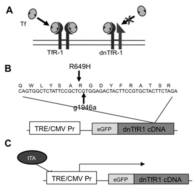
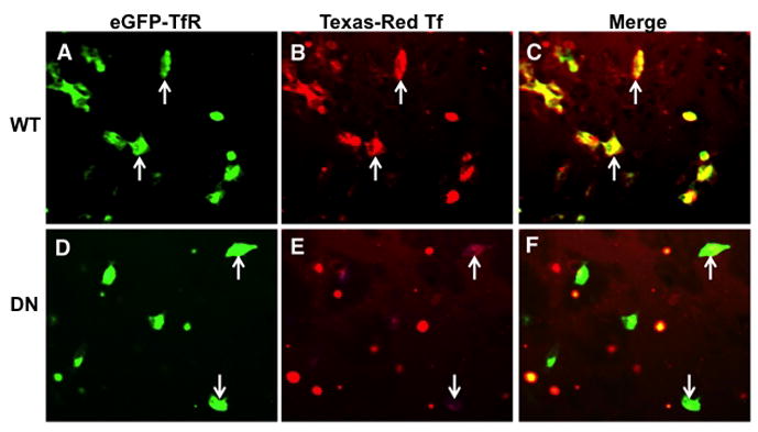
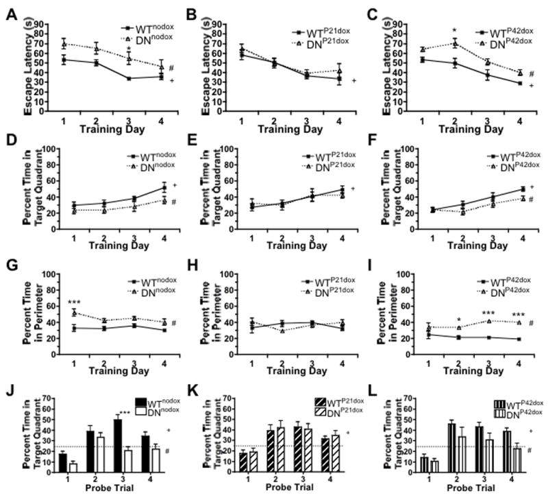
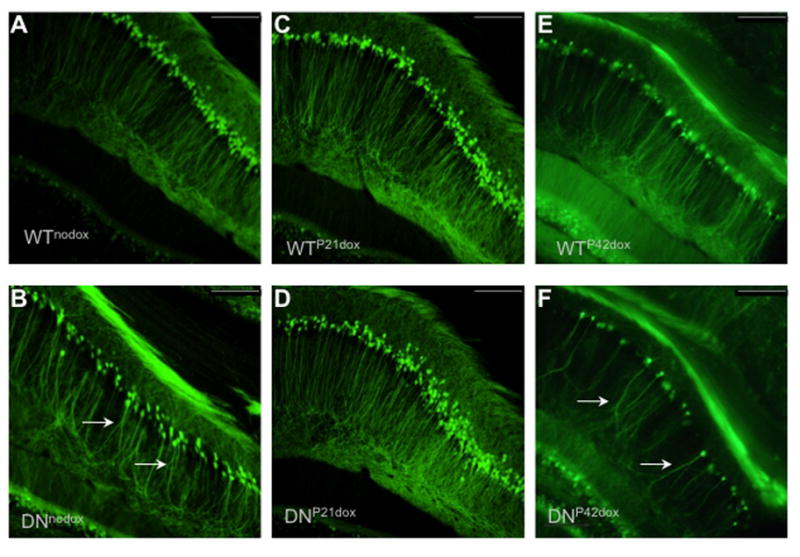
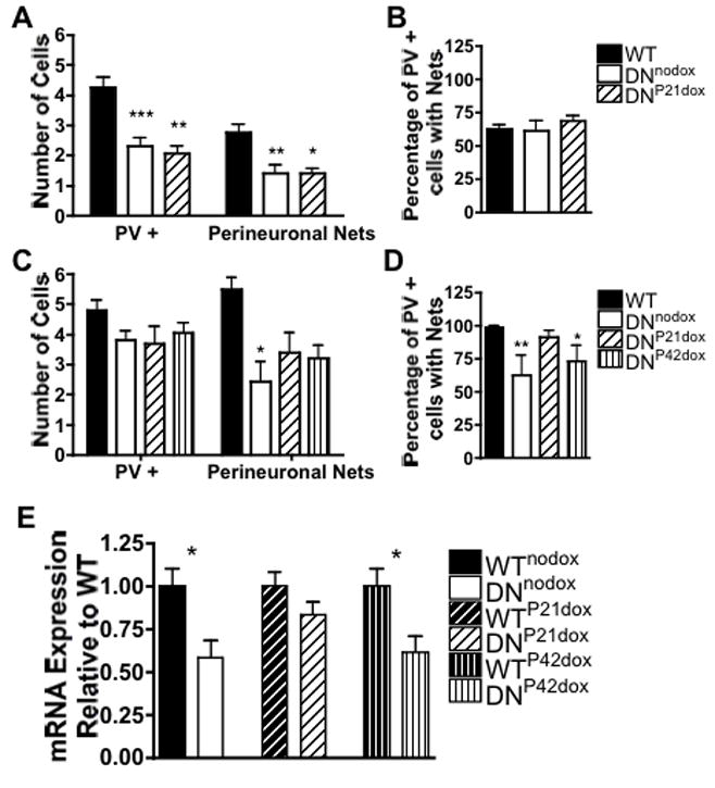
Iron is a necessary substrate for neuronal function throughout the lifespan, but particularly during development. Early life iron deficiency (ID) in humans (late gestation through 2-3 yr) results in persistent cognitive and behavioral abnormalities despite iron repletion. Animal models of early life ID generated using maternal dietary iron restriction also demonstrate persistent learning and memory deficits, suggesting a critical requirement for iron during hippocampal development. Precise definition of the temporal window for this requirement has been elusive due to anemia and total body and brain ID inherent to previous dietary restriction models. To circumvent these confounds, we developed transgenic mice that express tetracycline transactivator regulated, dominant negative transferrin receptor (DNTfR1) in hippocampal neurons, disrupting TfR1 mediated iron uptake specifically in CA1 pyramidal neurons. Normal iron status was restored by doxycycline administration. We manipulated the duration of ID using this inducible model to examine long-term effects of early ID on Morris water maze learning, CA1 apical dendrite structure, and defining factors of critical periods including parvalbmin (PV) expression, perineuronal nets (PNN), and brain-derived neurotrophic factor (BDNF) expression. Ongoing ID impaired spatial memory and resulted in disorganized apical dendrite structure accompanied by altered PV and PNN expression and reduced BDNF levels. Iron repletion at P21, near the end of hippocampal dendritogenesis, restored spatial memory, dendrite structure, and critical period markers in adult mice. However, mice that remained hippocampally iron deficient until P42 continued to have spatial memory deficits, impaired CA1 apical dendrite structure, and persistent alterations in PV and PNN expression and reduced BDNF despite iron repletion. Together, these findings demonstrate that hippocampal iron availability is necessary between P21 and P42 for development of normal spatial learning and memory, and that these effects may reflect disruption of critical period closure by early life ID.
Copyright © 2011 Wiley Periodicals, Inc.
Figures

Similar articles
-
Early-Life Neuronal-Specific Iron Deficiency Alters the Adult Mouse Hippocampal Transcriptome.J Nutr. 2018 Oct 1;148(10):1521-1528. doi: 10.1093/jn/nxy125. J Nutr. 2018. PMID: 30169712 Free PMC article.
-
Neuronal-specific iron deficiency dysregulates mammalian target of rapamycin signaling during hippocampal development in nonanemic genetic mouse models.J Nutr. 2013 Mar;143(3):260-6. doi: 10.3945/jn.112.168617. Epub 2013 Jan 9. J Nutr. 2013. PMID: 23303869 Free PMC article.
-
Early alterations in hippocampal perisomatic GABAergic synapses and network oscillations in a mouse model of Alzheimer's disease amyloidosis.PLoS One. 2019 Jan 15;14(1):e0209228. doi: 10.1371/journal.pone.0209228. eCollection 2019. PLoS One. 2019. PMID: 30645585 Free PMC article.
-
The role of iron in learning and memory.Adv Nutr. 2011 Mar;2(2):112-21. doi: 10.3945/an.110.000190. Epub 2011 Mar 10. Adv Nutr. 2011. PMID: 22332040 Free PMC article. Review.
-
The role of iron in neurodevelopment: fetal iron deficiency and the developing hippocampus.Biochem Soc Trans. 2008 Dec;36(Pt 6):1267-71. doi: 10.1042/BST0361267. Biochem Soc Trans. 2008. PMID: 19021538 Free PMC article. Review.
Cited by
-
An Extracellular Perspective on CNS Maturation: Perineuronal Nets and the Control of Plasticity.Int J Mol Sci. 2021 Feb 28;22(5):2434. doi: 10.3390/ijms22052434. Int J Mol Sci. 2021. PMID: 33670945 Free PMC article. Review.
-
Metabolomic analysis of CSF indicates brain metabolic impairment precedes hematological indices of anemia in the iron-deficient infant monkey.Nutr Neurosci. 2018 Jan;21(1):40-48. doi: 10.1080/1028415X.2016.1217119. Epub 2016 Aug 6. Nutr Neurosci. 2018. PMID: 27499134 Free PMC article.
-
Early life nutrition and neural plasticity.Dev Psychopathol. 2015 May;27(2):411-23. doi: 10.1017/S0954579415000061. Dev Psychopathol. 2015. PMID: 25997762 Free PMC article. Review.
-
The importance of iron deficiency in pregnancy on fetal, neonatal, and infant neurodevelopmental outcomes.Int J Gynaecol Obstet. 2023 Aug;162 Suppl 2(Suppl 2):83-88. doi: 10.1002/ijgo.14951. Int J Gynaecol Obstet. 2023. PMID: 37538010 Free PMC article. Review.
-
Multi-omic analysis characterizes molecular susceptibility of receptors to SARS-CoV-2 spike protein.Comput Struct Biotechnol J. 2023 Nov 10;21:5583-5600. doi: 10.1016/j.csbj.2023.11.012. eCollection 2023. Comput Struct Biotechnol J. 2023. PMID: 38034398 Free PMC article.
References
-
- Bekenstein JW, Lothman EW. A comparison of the ontogeny of excitatory and inhibitory neurotransmission in the CA1 region and dentate gyrus of the rat hippocampal formation. Brain Res Dev Brain Res. 1991a;63(1–2):237–43. - PubMed
-
- Bekenstein JW, Lothman EW. An in vivo study of the ontogeny of long-term potentiation (LTP) in the CA1 region and in the dentate gyrus of the rat hippocampal formation. Brain Res Dev Brain Res. 1991b;63(1–2):245–51. - PubMed
Publication types
MeSH terms
Substances
Grants and funding
LinkOut - more resources
Full Text Sources
Medical
Molecular Biology Databases
Miscellaneous
