

Therapeutic devices for epilepsy
- PMID: 22367987
- PMCID: PMC3296971
- DOI: 10.1002/ana.22621
Therapeutic devices for epilepsy
Abstract
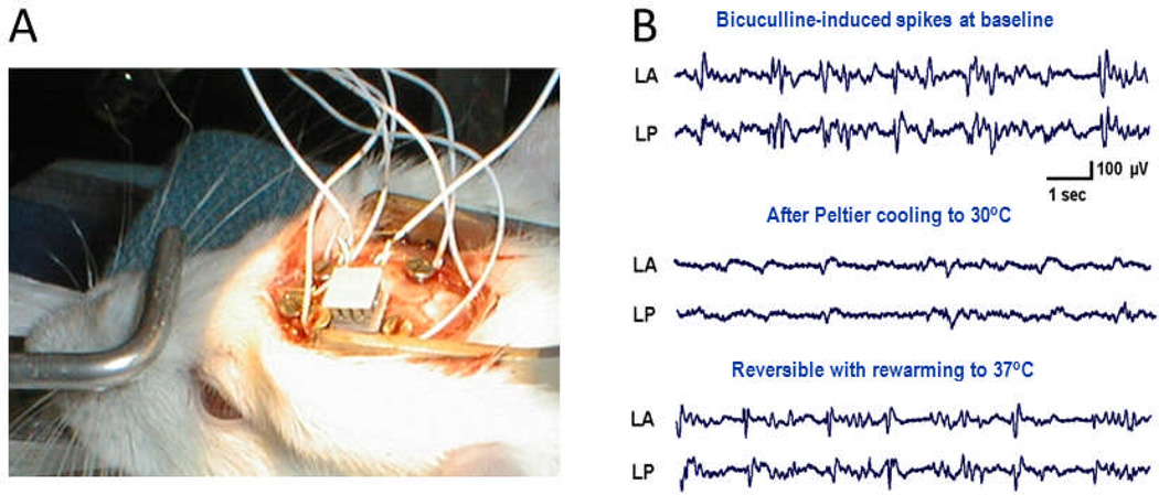
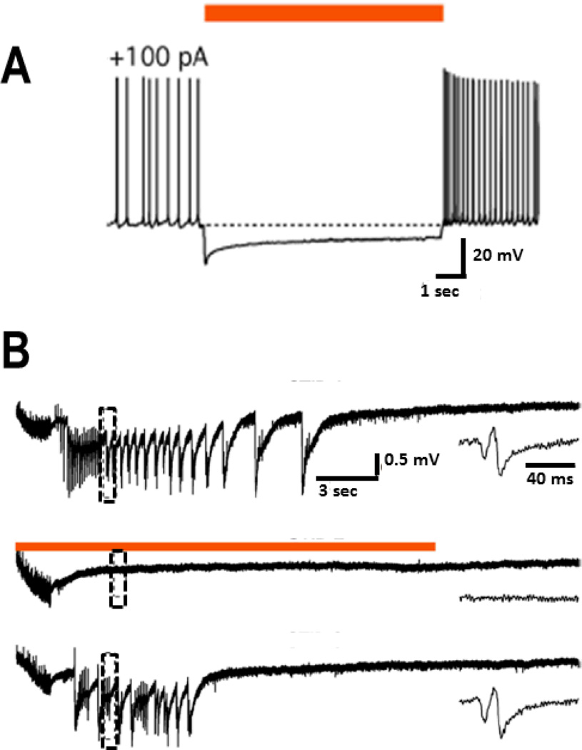
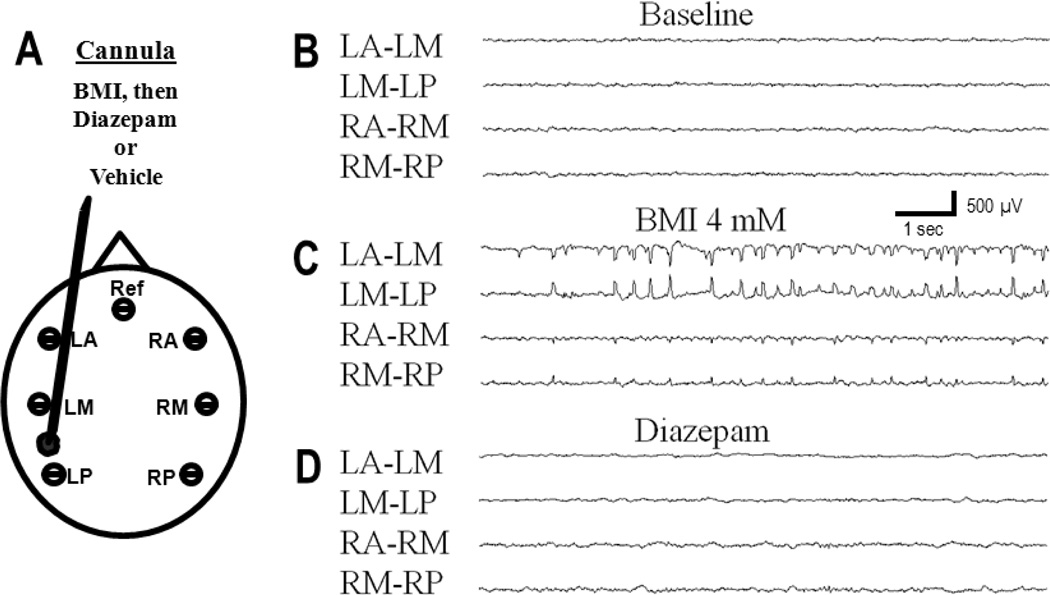
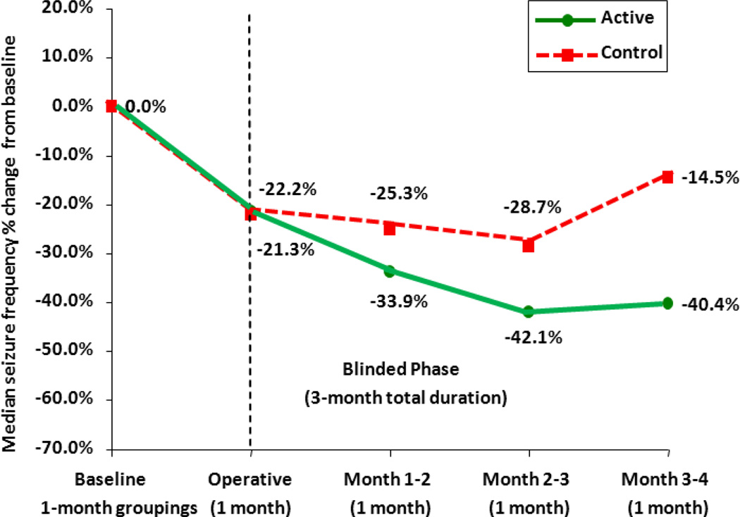
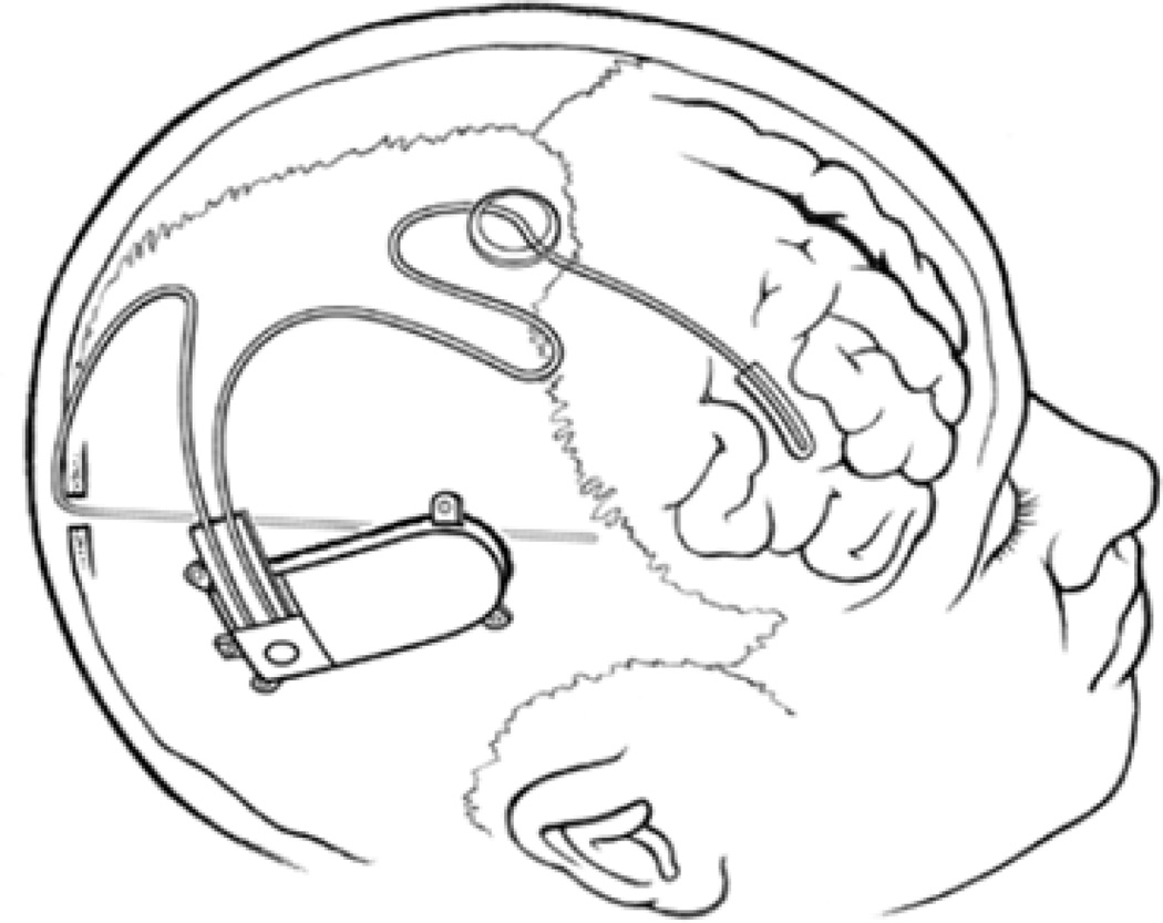
Therapeutic devices provide new options for treating drug-resistant epilepsy. These devices act by a variety of mechanisms to modulate neuronal activity. Only vagus nerve stimulation (VNS), which continues to develop new technology, is approved for use in the United States. Deep brain stimulation of anterior thalamus for partial epilepsy recently was approved in Europe and several other countries. Responsive neurostimulation, which delivers stimuli to 1 or 2 seizure foci in response to a detected seizure, recently completed a successful multicenter trial. Several other trials of brain stimulation are in planning or underway. Transcutaneous magnetic stimulation (TMS) may provide a noninvasive method to stimulate cortex. Controlled studies of TMS are split on efficacy, which may depend on whether a seizure focus is near a possible region for stimulation. Seizure detection devices in the form of shake detectors via portable accelerometers can provide notification of an ongoing tonic-clonic seizure, or peace of mind in the absence of notification. Prediction of seizures from various aspects of electroencephalography (EEG) is in early stages. Prediction appears to be possible in a subpopulation of people with refractory seizures, and a clinical trial of an implantable prediction device is underway. Cooling of neocortex or hippocampus reversibly can attenuate epileptiform EEG activity and seizures, but engineering problems remain in its implementation. Optogenetics is a new technique that can control excitability of specific populations of neurons with light. Inhibition of epileptiform activity has been demonstrated in hippocampal slices, but use in humans will require more work. In general, devices provide useful palliation for otherwise uncontrollable seizures, but with a different risk profile than with most drugs. Optimizing the place of devices in therapy for epilepsy will require further development and clinical experience.
Copyright © 2011 American Neurological Association.
Figures
References
-
- Carlson C, Arnedo V, Cahill M, Devinsky O. Detecting nocturnal convulsions: efficacy of the MP5 monitor. Seizure. 2009;18:225–227. - PubMed
-
- Karayiannis NB, Xiong Y, Frost JD, Jr, Wise MS, Hrachovy RA, Mizrahi EM. Automated detection of videotaped neonatal seizures based on motion tracking methods. J Clin Neurophysiol. 2006;23:521–531. - PubMed
-
- Gotman J. Automatic recognition of epileptic seizures in the EEG. Electroencephalogr Clin Neurophysiol. 1982;54:530–540. - PubMed
-
- Elzawahry H, Do CS, Lin K, Benbadis SR. The diagnostic utility of the ictal cry. Epilepsy Behav. 2010;18:306–307. - PubMed
-
- Leutmezer F, Schernthaner C, Lurger S, Potzelberger K, Baumgartner C. Electrocardiographic changes at the onset of epileptic seizures. Epilepsia. 2003;44:348–354. - PubMed
Publication types
MeSH terms
Grants and funding
LinkOut - more resources
Full Text Sources
Other Literature Sources
Medical
