

High-frequency oscillations as a new biomarker in epilepsy
- PMID: 22367988
- PMCID: PMC3754947
- DOI: 10.1002/ana.22548
High-frequency oscillations as a new biomarker in epilepsy
Abstract
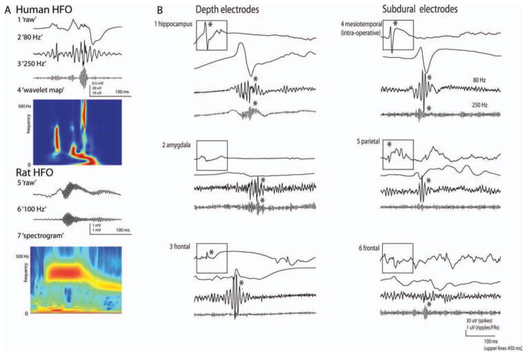
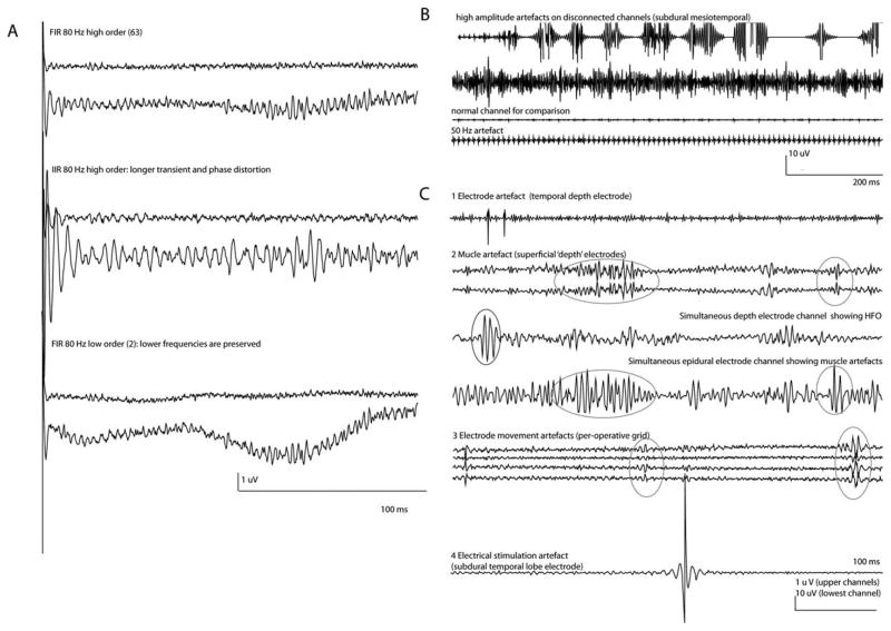
The discovery that electroencephalography (EEG) contains useful information at frequencies above the traditional 80Hz limit has had a profound impact on our understanding of brain function. In epilepsy, high-frequency oscillations (HFOs, >80Hz) have proven particularly important and useful. This literature review describes the morphology, clinical meaning, and pathophysiology of epileptic HFOs. To record HFOs, the intracranial EEG needs to be sampled at least at 2,000Hz. The oscillatory events can be visualized by applying a high-pass filter and increasing the time and amplitude scales, or EEG time-frequency maps can show the amount of high-frequency activity. HFOs appear excellent markers for the epileptogenic zone. In patients with focal epilepsy who can benefit from surgery, invasive EEG is often required to identify the epileptic cortex, but current information is sometimes inadequate. Removal of brain tissue generating HFOs has been related to better postsurgical outcome than removing the seizure onset zone, indicating that HFOs may mark cortex that needs to be removed to achieve seizure control. The pathophysiology of epileptic HFOs is challenging, probably involving populations of neurons firing asynchronously. They differ from physiological HFOs in not being paced by rhythmic inhibitory activity and in their possible origin from population spikes. Their link to the epileptogenic zone argues that their study will teach us much about the pathophysiology of epileptogenesis and ictogenesis. HFOs show promise for improving surgical outcome and accelerating intracranial EEG investigations. Their potential needs to be assessed by future research.
Copyright © 2012 American Neurological Association.
Conflict of interest statement
P.J. has received grant(s) and has grants/grants pending from Epilepsy Research UK. J.G.R.J. has received grant(s) from the Medical Research Council (UK) and Epilepsy Research UK, and has received support for travel to the European Epilepsy Congress from UCB. M.Z. has received grant(s) from the Netherlands Organization for Scientific Research. J.G. has received grant(s) from the Canadian Institutes of Health Research; has had consultancies with Blackrock Microsystems; and has been employed by Stellate Systems. R.Z. has received grant(s) from the Natural Sciences and Engineering Research Council of Canada (NSERC-PGSD) and the Canadian Institutes of Health Research (CIHR).
Figures
References
-
- Allen PJ, Fish DR, Smith SJ. Very high-frequency rhythmic activity during SEEG suppression in frontal lobe epilepsy. Electroencephalogr Clin Neurophysiol. 1992;82:155–159. - PubMed
-
- Fisher RS, Webber WR, Lesser RP, et al. High-frequency EEG activity at the start of seizures. J Clin Neurophysiol. 1992;9:441–448. - PubMed
-
- Huang CM, White LE., Jr High-frequency components in epileptiform EEG. J Neurosci Methods. 1989;30:197–201. - PubMed
-
- Bragin A, Engel J, Jr, Wilson CL, et al. Electrophysiologic analysis of a chronic seizure model after unilateral hippocampal KA injection. Epilepsia. 1999;40:1210–1221. - PubMed
-
- Bragin A, Engel J, Jr, Wilson CL, et al. Hippocampal and entorhinal cortex high-frequency oscillations (100–500 Hz) in human epileptic brain and in kainic acid–treated rats with chronic seizures. Epilepsia. 1999;40:127–137. - PubMed
Publication types
MeSH terms
Grants and funding
LinkOut - more resources
Full Text Sources
Other Literature Sources
Medical
