

Wide variation and rising utilization of stroke magnetic resonance imaging: data from 11 states
- PMID: 22367989
- PMCID: PMC3297973
- DOI: 10.1002/ana.22698
Wide variation and rising utilization of stroke magnetic resonance imaging: data from 11 states
Abstract
Objective: Neuroimaging is an essential component of the acute stroke evaluation. Magnetic resonance imaging (MRI) is more accurate than computed tomography (CT) for the diagnosis of stroke, but is more costly and time-consuming. We sought to describe changes in MRI utilization from 1999 to 2008.
Methods: We performed a serial cross-sectional study with time trends of neuroimaging in patients with a primary International Classification of Diseases, 9th Edition, Clinical Modification discharge diagnosis of stroke admitted through the emergency department in the State Inpatient Databases from 10 states. MRI utilization was measured by Healthcare Cost and Utilization Project criteria. Data were included for states from 1999 to 2008 where MRI utilization could be identified.
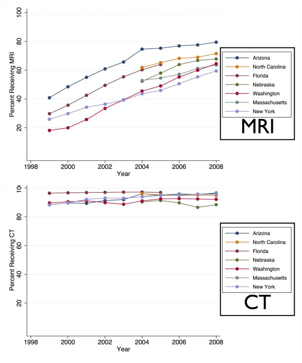
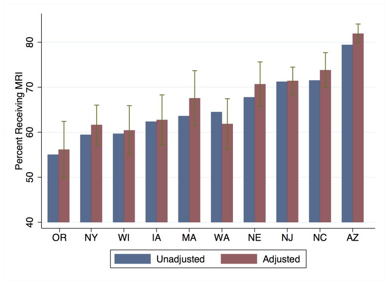
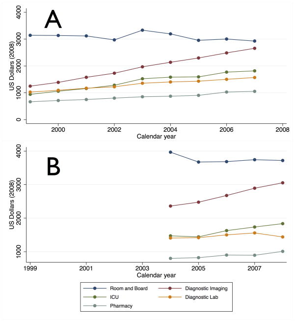
Results: A total of 624,842 patients were hospitalized for stroke in the period of interest. MRI utilization increased in all states. Overall, MRI absolute utilization increased 38%, and relative utilization increased 235% (28% of strokes in 1999 to 66% in 2008). Over the same interval, CT utilization changed little (92% in 1999 to 95% in 2008). MRI use varied widely by state. In 2008, MRI utilization ranged from a low of 55% of strokes in Oregon to a high of 79% in Arizona. Diagnostic imaging was the fastest growing component of total hospital costs (213% increase from 1999 to 2007).
Interpretation: MRI utilization during stroke hospitalization increased substantially, with wide geographic variation. Rather than replacing CT, MRI is supplementing it. Consequently, neuroimaging has been the fastest growing component of hospitalization cost in stroke. Recent neuroimaging practices in stroke are not standardized and may represent an opportunity to improve the efficiency of stroke care.
Copyright © 2012 American Neurological Association.
Figures
Comment in
-
Modern care for neurological problems must address waste.Ann Neurol. 2012 Feb;71(2):A5-6. doi: 10.1002/ana.23539. Ann Neurol. 2012. PMID: 22368006 No abstract available.
References
-
- Brazzelli M, Sandercock PA, Chappell FM, et al. Magnetic resonance imaging versus computed tomography for detection of acute vascular lesions in patients presenting with stroke symptoms. Cochrane Database Syst Rev. 2009 Jan;1(4):CD007424. - PubMed
-
- Barber PA, Darby DG, Desmond PM, et al. Identification of major ischemic change. Diffusion-weighted imaging versus computed tomography. Stroke; a journal of cerebral circulation. 1999 Oct 01;30(10):2059–65. - PubMed
-
- Fiebach JB, Schellinger PD, Jansen O, et al. CT and diffusion-weighted MR imaging in randomized order: diffusion-weighted imaging results in higher accuracy and lower interrater variability in the diagnosis of hyperacute ischemic stroke. Stroke; a journal of cerebral circulation. 2002 Sep 01;33(9):2206–10. - PubMed
-
- González RG, Schaefer PW, Buonanno FS, et al. Diffusion-weighted MR imaging: diagnostic accuracy in patients imaged within 6 hours of stroke symptom onset. Radiology. 1999;210(1):155–62. - PubMed
