. 2012 May;71(5):687-98.
doi: 10.1002/ana.23536. Epub 2012 Feb 24.

Anesthetics isoflurane and desflurane differently affect mitochondrial function, learning, and memory

Affiliations

Anesthetics isoflurane and desflurane differently affect mitochondrial function, learning, and memory

Yiying Zhang et al. Ann Neurol. 2012 May.

Abstract

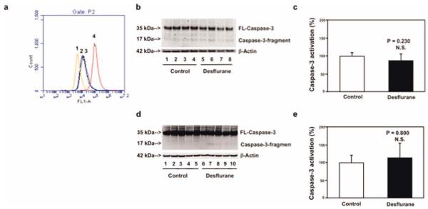
Objective: There are approximately 8.5 million Alzheimer disease (AD) patients who need anesthesia and surgery care every year. The inhalation anesthetic isoflurane, but not desflurane, has been shown to induce caspase activation and apoptosis, which are part of AD neuropathogenesis, through the mitochondria-dependent apoptosis pathway. However, the in vivo relevance, underlying mechanisms, and functional consequences of these findings remain largely to be determined.

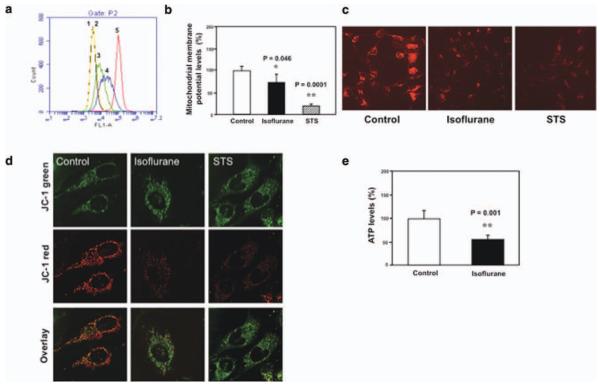
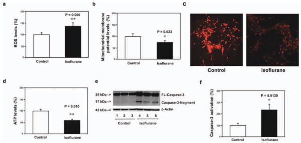
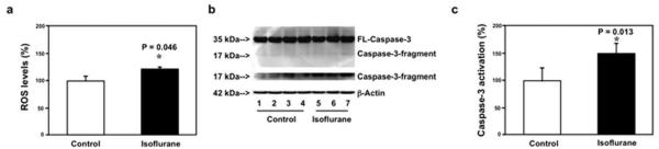
Methods: We therefore set out to assess the effects of isoflurane and desflurane on mitochondrial function, cytotoxicity, learning, and memory using flow cytometry, confocal microscopy, Western blot analysis, immunocytochemistry, and the fear conditioning test.

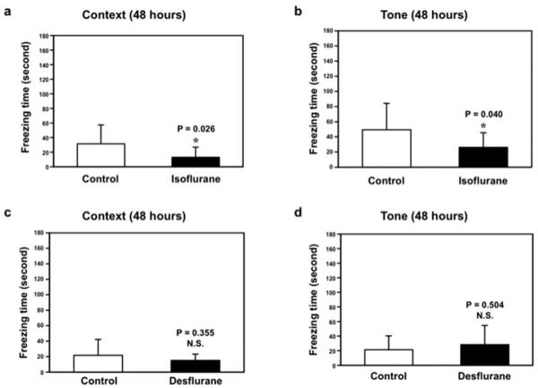
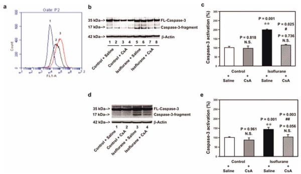
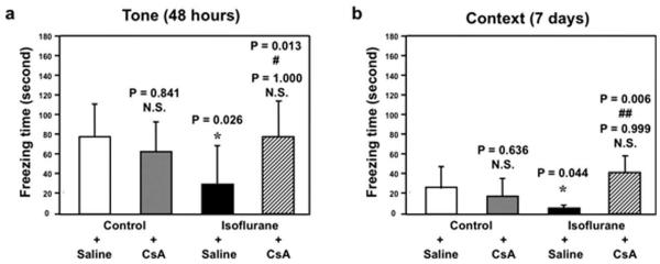
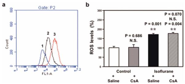
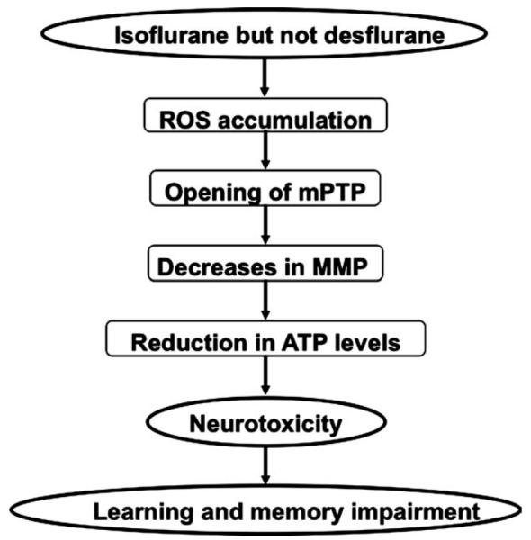
Results: Here we show that isoflurane, but not desflurane, induces opening of mitochondrial permeability transition pore (mPTP), increase in levels of reactive oxygen species, reduction in levels of mitochondrial membrane potential and adenosine-5'-triphosphate, activation of caspase 3, and impairment of learning and memory in cultured cells, mouse hippocampus neurons, mouse hippocampus, and mice. Moreover, cyclosporine A, a blocker of mPTP opening, attenuates isoflurane-induced mPTP opening, caspase 3 activation, and impairment of learning and memory. Finally, isoflurane may induce the opening of mPTP via increasing levels of reactive oxygen species.

Interpretation: These findings suggest that desflurane could be a safer anesthetic for AD patients as compared to isoflurane, and elucidate the potential mitochondria-associated underlying mechanisms, and therefore have implications for use of anesthetics in AD patients, pending human study confirmation.


Figures

FIGURE 1
FIGURE 1
Isoflurane induces opening of mitochondrial permeability transition pore (mPTP), and decreases levels of mitochondrial membrane potential (MMP) and adenosine-5′-triphosphate (ATP) in B104 cells and H4-APP cells. (a) Flow cytometric analysis shows changes in calcein levels in mitochondria of B104 cells stained with calcein AM or calcein AM plus cobalt, which indicates opening of mPTP: peak 1, unstained B104 cells; peak 2, positive control (treatment of calcein AM plus cobalt and ionomycin); peak 3, cells treated with calcein AM plus cobalt and isoflurane; peak 4, negative control (treatment of calcein AM plus cobalt); peak 5, calcein AM treated B104 cells. The changes in intensity of fluorescence between isoflurane group (peak 3), positive control (peak 2), and negative control (peak 4) suggest that isoflurane induces opening of mPTP. (b) Tetraethylbenzimidazolylcarbocyanine iodide (JC-1) fluorescence analysis shows that isoflurane (black bar) or staurosporine (STS; net bar) reduces levels of MMP as compared to control condition. (c) Isoflurane or STS may decrease levels of MMP, detected by staining of tetramethylrhodamine ethyl ester and perchlorate, the MMP-dependent fluorescent indicator, as compared to control condition. (d) Isoflurane or STS may decrease levels of MMP, detected by JC-1 staining, as compared to control condition. The first row illustrates that there is no significant difference of JC-1 green staining following treatments of control condition, isoflurane, and STS. The second row illustrates that isoflurane or STS may decrease MMP, detected by JC-1 red staining, as compared to control condition. The third row is an overlay of JC-1 green and JC-1 red staining. (e) Isoflurane reduces ATP levels (black bar) as compared to control condition (white bar).
FIGURE 2
FIGURE 2
Isoflurane increases reactive oxygen species (ROS) levels, decreases levels of mitochondrial membrane potential (MMP) and adenosine-5′-triphosphate (ATP), and induces caspase 3 activation in mouse hippocampus neurons. (a) Isoflurane (black bar) increases ROS levels as compared to the control condition (white bar) in mouse hippocampus neurons. (b) Tetraethylbenzimidazolylcarbocyanine iodide fluorescence analysis shows that isoflurane (black bar) reduces levels of MMP as compared to the control condition in mouse hippocampus neurons. (v) Isoflurane may decrease levels of MMP, detected by staining of tetramethylrhodamine ethyl ester and perchlorate, the MMP-dependent fluorescent indicator, as compared to the control condition in mouse hippocampus neurons. (d) Isoflurane (black bar) reduces ATP levels as compared to the control condition (white bar) in mouse hippocampus neurons. (e) Western blot shows that isoflurane (lanes 4–6) induces caspase 3 activation as compared to the control condition (lanes 1–3) in mouse hippocampus neurons. There is no significant difference in the amounts of β-actin in the control condition or the isoflurane-treated mouse hippocampus neurons. (f) Quantification of the Western blot shows that isoflurane (black bar) induces caspase 3 activation as compared to the control condition (white bar) in mouse hippocampus neurons. FL = full length.
FIGURE 3
FIGURE 3
Isoflurane increases reactive oxygen species (ROS) levels and induces caspase 3 activation in mouse hippocampus. (a) Isoflurane (black bar) increases ROS levels as compared to the control condition (white bar) in mouse hippocampus. (b) Western blot shows that isoflurane (lanes 5–7) induces caspase 3 activation as compared to the control condition (lanes 1–4) in mouse hippocampus. There is no significant difference in amounts of β-actin in the control condition or isoflurane-treated mice. (c) Quantification of the Western blot shows that isoflurane (black bar) induces caspase 3 activation as compared to the control condition (white bar) in mouse hippocampus. FL = full length.
FIGURE 4
FIGURE 4
Desflurane induces neither opening of mitochondrial permeability transition pore (mPTP) nor caspase 3 activation in B104 cells and mouse brain tissues. (a) Flow cytometric analysis shows changes in calcein levels in mitochondria of B104 cells stained with calcein AM or calcein AM plus cobalt, which indicates opening of mPTP: peak 1, treatment of calcein AM plus cobalt and ionomycin (positive control of opening of mPTP); peak 2, treatment of calcein AM plus cobalt and desflurane; peak 3, treatment of calcein AM plus cobalt (negative control); peak 4, treatment of calcein AM. The position of desflurane treatment (peak 2) locates away from that of positive control (peak 1) but overlaps with that of negative control (peak 3), which suggests that desflurane may not induce opening of mPTP. (b) Treatment with 12% desflurane for 6 hours (lanes 5–8) does not induce caspase 3 activation as compared to control condition (lanes 1–4) in B104 cells. There is no significant difference in amounts of β-actin in control condition or desflurane-treated B104 cells. (c) Quantification of the Western blot shows that desflurane (black bar) does not induce caspase 3 activation as compared to control condition (white bar) in B104 cells. (d) Treatment of 7.5% desflurane for 6 hours (lanes 6–10) does not induce caspase 3 activation in brain tissues of mice as compared to control condition (lanes 1–5). There is no significant difference in amounts of β-actin in control condition or desflurane-treated mouse brain tissues. (E) Quantification of the Western blot shows that desflurane (black bar) does not induce caspase 3 activation as compared to control condition (white bar) in mouse brain tissues. FL = full length; N.S. = not significant. [Color figure can be viewed in the online issue, which is available at annalsofneurology.org.]
FIGURE 5
FIGURE 5
Isoflurane, but not desflurane, induces learning and memory impairment in mice. (a) Isoflurane (black bar) decreases freezing time in the context test of the fear conditioning test (FCT) as compared to control condition (white bar) at 48 hours after isoflurane treatment. (b) Isoflurane (black bar) decreases freezing time in the tone test of the FCT as compared to control condition (white bar) at 48 hours after isoflurane anesthesia. Desflurane does not decrease freezing time in the (c) context test and (D) tone test of the FCT at 48 hours after desflurane anesthesia. N.S. = not significant.
FIGURE 6
FIGURE 6
Cyclosporine A (CsA) attenuates isoflurane-induced opening of mitochondrial permeability transition pore (mPTP), caspase 3 activation in B104 cells and brain tissues of mice. (a) Flow cytometric analysis shows changes in calcein levels in mitochondria of B104 cells stained with calcein AM or calcein AM plus cobalt, which indicates opening of mPTP: peak 1, treatment of ionomycin (the positive control of opening of mPTP); peak 2, treatment of isoflurane; peak 3, treatment of isoflurane plus CsA (1μM). CsA treatment attenuates isoflurane-induced opening of mPTP, as demonstrated by the position of peak of isoflurane treatment shifting to the right following CsA treatment. (b) Western blot shows that treatment of 2% isoflurane for 6 hours (lanes 5 and 6) induces caspase 3 activation as compared to control condition (lanes 1 and 2) in B104 cells. CsA treatment alone (lanes 3 and 4) does not induce caspase 3 activation as compared to control condition (lanes 1 and 2), but CsA treatment attenuates isoflurane-induced caspase 3 activation (lanes 7 and 8) as compared to isoflurane treatment (lanes 5 and 6). (c) Quantification of the Western blot shows that isoflurane (black bar) induces caspase 3 activation as compared to control condition (white bar). CsA treatment (net bar) attenuates isoflurane-induced caspase 3 activation as compared to isoflurane treatment (black bar) in B104 cells. (d) Western blot shows that treatment of 1.4% isoflurane for 6 hours (lane 3) induces caspase 3 activation as compared to control condition (lane 1). CsA treatment alone (lane 2) does not induce caspase 3 activation as compared to control condition (lane 1), but CsA treatment attenuates isoflurane-induced caspase 3 activation (lane 4) as compared to isoflurane treatment (lane 3). (e) Quantification of the Western blot shows that isoflurane (black bar) induces caspase 3 activation as compared to control condition (white bar). CsA treatment (net bar) attenuates isoflurane-induced caspase 3 activation as compared to isoflurane treatment (black bar) in mouse brain tissues. FL = full length; N.S. = not significant. [Color figure can be viewed in the online issue, which is available at annalsofneurology.org.]
FIGURE 7
FIGURE 7
Cyclosporine A (CsA) attenuates isoflurane-induced learning and memory impairment in mice. (a) Isoflurane treatment (black bar) decreases freezing time in the tone test of the fear conditioning test (FCT) as compared to control condition (white bar) at 48 hours after isoflurane anesthesia. CsA treatment alone (gray bar) does not significantly affect freezing time as compared to control condition (white bar) in the tone test of the FCT at 48 hours after the treatment. However, 2-way analysis of variance (ANOVA) shows that there is an interaction of CsA and isoflurane in that CsA treatment (net bar) attenuates isoflurane-induced reduction in freezing time in the tone test of the FCT at 48 hours after the treatment. (b) Isoflurane treatment (black bar) decreases freezing time in the context test of the FCT as compared to control condition (white bar) at 7 days after isoflurane anesthesia. CsA treatment alone (gray bar) does not significantly affect freezing time as compared to control condition (white bar) in the context test of the FCT at 7 days after the treatment. However, 2-way ANOVA shows that there is an interaction of CsA and isoflurane in that CsA treatment (net bar) attenuates isoflurane-induced reduction in freezing time in the context test of the FCT at 7 days after the treatment. N.S. = not significant.
FIGURE 8
FIGURE 8
Effects of N-acetyl-L-cysteine (NAC) and cyclosporine A (CsA) on isoflurane-induced opening of mitochondrial permeability transition pore (mPTP) and increases in reactive oxygen species (ROS) levels. (a) Flow cytometric analysis shows changes in calcein levels in mitochondria of B104 cells stained with calcein AM or calcein AM plus cobalt, which indicates opening of mPTP: peak 1, treatment of ionomycin (positive control of opening of mPTP); peak 2, isoflurane treatment; peak 3, treatment of isoflurane plus NAC (1mM), the inhibitor of ROS generation. NAC treatment attenuates isoflurane-induced opening of mPTP, as demonstrated by the position of the peak of isoflurane treatment shifting to the right following NAC treatment in B104 cells. (b) Isoflurane treatment (black bar) increases ROS levels as compared to control condition (white bar) in H4-APP cells. CsA treatment alone (gray bar) does not decrease ROS levels as compared to control condition (white bar); 2-way analysis of variance shows that there is no interaction of isoflurane and CsA on ROS levels in that CsA treatment (net bar) does not attenuate isoflurane-induced increases of ROS levels. N.S. = not significant. [Color figure can be viewed in the online issue, which is available at annalsofneurology.org.]
FIGURE 9
FIGURE 9
Hypothetical pathway by which isoflurane induces cytotoxicity. Isoflurane, but not desflurane, induces reactive oxygen species (ROS) accumulation, which then facilitates opening of mitochondrial permeability transition pore (mPTP). Opening of mPTP will cause decreases in levels of mitochondrial membrane potential (MMP), and consequently reduction in adenosine-5′-triphosphate (ATP) levels, leading to neurotoxicity (eg, caspase 3 activation) and finally impairment of learning and memory.

References

    1. Alzheimer’s Association . Alzheimer’s Disease Facts and Figures. Alzheimer’s Association; Chicago, IL: 2011. http://www.alz.org/downloads/Facts_Figures_2011.pdf. - PubMed
    1. Silbert B, Evered L, Scott DA, Maruff P. Anesthesiology must play a greater role in patients with Alzheimer’s disease. Anesth Analg. 2011;112:1242–1245. - PubMed
    1. Bittner EA, Yue Y, Xie Z. Brief review: anesthetic neurotoxicity in the elderly, cognitive dysfunction and Alzheimer’s disease. Can J Anaesth. 2011;58:216–223. - PMC - PubMed
    1. Baranov D, Bickler PE, Crosby GJ, et al. Consensus statement: First International Workshop on Anesthetics and Alzheimer’s disease. Anesth Analg. 2009;108:1627–1630. - PMC - PubMed
    1. Eckenhoff RG, Johansson JS, Wei H, et al. Inhaled anesthetic enhancement of amyloid-beta oligomerization and cytotoxicity. Anesthesiology. 2004;101:703–709. - PubMed

Publication types

MeSH terms

Substances