Comparison between scalpel technique and electrosurgery for depigmentation: A case series
- PMID: 22368368
- PMCID: PMC3283941
- DOI: 10.4103/0972-124X.92580
Comparison between scalpel technique and electrosurgery for depigmentation: A case series
Abstract
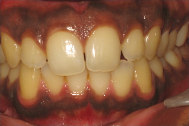
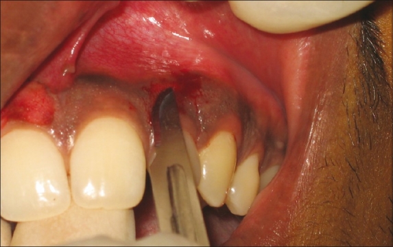
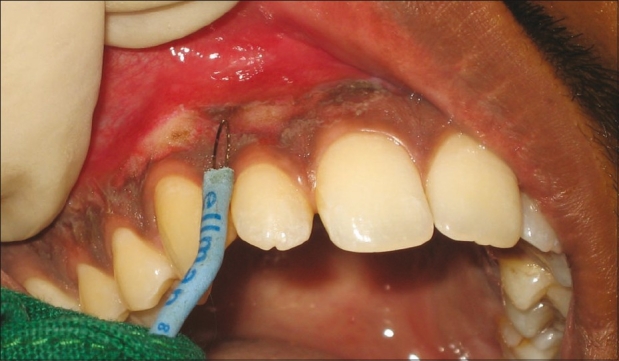
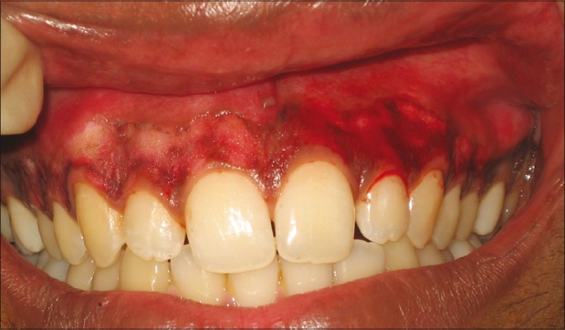
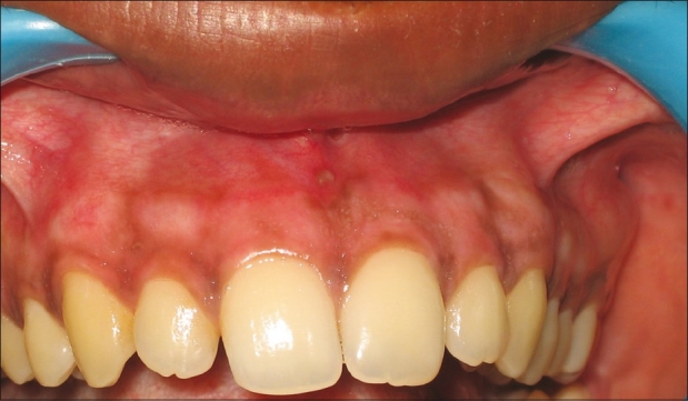
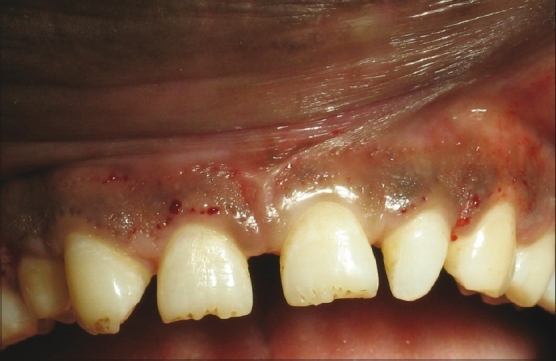
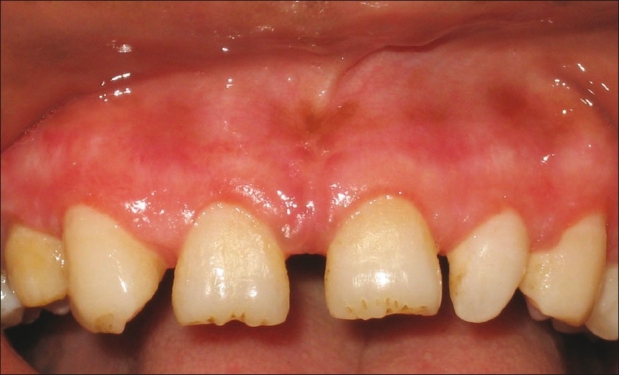
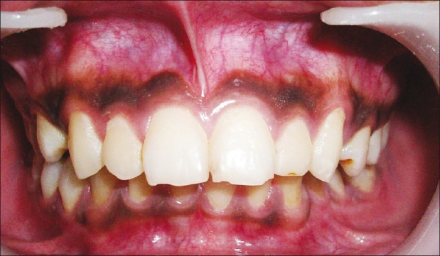
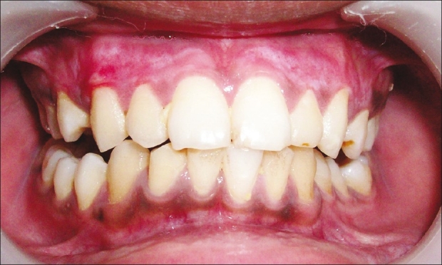
A beautiful smile definitely enhances the personality of an individual and reveals self-confidence. The harmony of the smile is determined not only by the shape, position, and color of the teeth but also by the gingival tissues. Gingival pigmentation results from melanin granules which are produced by melanoblasts. Although melanin pigmentation of the gingiva is a completely benign condition and does not pose any medical problem, complaints of "black gums" are common particularly in patients having a very high smile line. The different treatment modalities that have been reported for depigmentation are bur abrasion, partial thickness flap, cryotherapy, electrosurgery, and lasers. In this paper we have compared the results of electrosurgery and scalpel technique, i.e., partial thickness flap.
Keywords: Depigmentation; electrosurgery; pigmented gingiva; scalpel technique.
Conflict of interest statement
Figures
References
-
- Dummett CO. Oral pigmentation. First symposium of oral pigmentation. J Periodontol. 1960;31:356–60.
-
- Cicek Y, Ertas U. The normal and pathological pigmentation of oral mucous membrane: A review. J Contemp Dent Pract. 2003;4:76–86. - PubMed
-
- Dummet CO, Barens G. Oromucosal pigmentation: An updated literary review. J Periodontol. 1971;42:726–36. - PubMed
-
- Perlmutter S, Tal H. Repigmentation of gingival following injury. J Periodontal. 1986;57:48–50. - PubMed
-
- Shafer WG, Hine MK, Levy BM. Philadelphia: WB Saunders Co; 1984. Text Book of Oral Pathology; pp. 89–136.