Review
. 2012;12(1):612-31.
doi: 10.3390/s120100612. Epub 2012 Jan 9.

Aptamers and their biological applications

Affiliations
Review

Aptamers and their biological applications

Kyung-Mi Song et al. Sensors (Basel). 2012.

Abstract

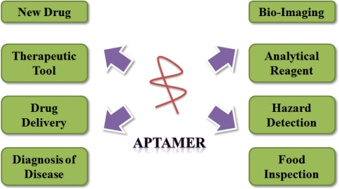
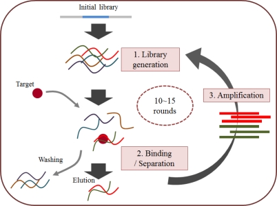
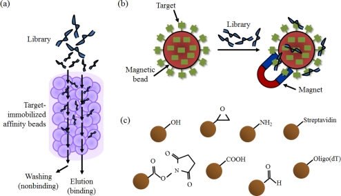
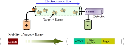
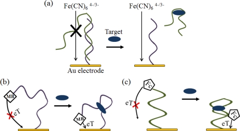
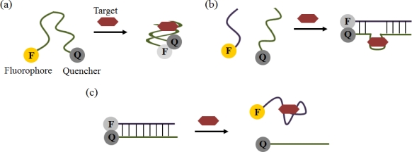
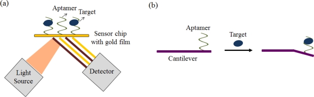
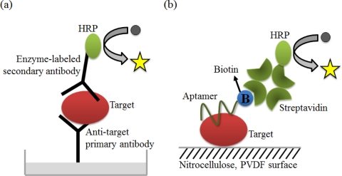
Recently, aptamers have attracted the attention of many scientists, because they not only have all of the advantages of antibodies, but also have unique merits, such as thermal stability, low cost, and unlimited applications. In this review, we present the reasons why aptamers are known as alternatives to antibodies. Furthermore, several types of in vitro selection processes, including nitrocellulose membrane filtration, affinity chromatography, magnetic bead, and capillary electrophoresis-based selection methods, are explained in detail. We also introduce various applications of aptamers for the diagnosis of diseases and detection of small molecules. Numerous analytical techniques, such as electrochemical, colorimetric, optical, and mass-sensitive methods, can be utilized to detect targets, due to convenient modifications and the stability of aptamers. Finally, several medical and analytical applications of aptamers are presented. In summary, aptamers are promising materials for diverse areas, not just as alternatives to antibodies, but as the core components of medical and analytical equipment.

Keywords: SELEX; aptamer; aptasensor; biosensor; diagnosis; in vitro selection.


Figures

Figure 1.
Figure 1.
Various application fields of aptamers.
Figure 2.
Figure 2.
The general SELEX strategy. Starting with combinatorial libraries (first step), the specific binders are isolated by an iterative process of ligand binding, elution (second step), and amplification (third step).
Figure 3.
Figure 3.
(a) A schematic illustration of the selection step from a library using an affinity column; (b) The process of the selection step using magnetic beads; (c) Several types of functional group-activated beads such as tosyl-activated beads and epoxy-activated beads.
Figure 4.
Figure 4.
Fundamental principle of the selection step using CE. Aptamers are selected based on the difference in mobility due to charge and mass.
Figure 5.
Figure 5.
Examples of electrochemical aptasensors. (a) A schematic representation of the electrochemical aptasensor using Fe(CN)64−/3−. Part of the aptamer was a hybridized aptamer with complementary DNA, which was immobilized on the gold surface. In the presence of the target, the aptamer was followed by binding with the target, to decrease the amount of the aptamer on the electrode surface; (b) A schematic representation of the electrochemical aptasensor using MB. In the presence of the target, the aptamer folds into the target-binding three-way junction, altering the electron transfer (eT) and increasing the observed reduction peak; (c) A schematic representation of the electrochemical aptasensor using Fc. In the presence of the target, the aptamer folds into the restricted hairpin structure, and this conformational change results in increased efficiency of eT between the Fc probe and the electrode surface.
Figure 6.
Figure 6.
Schematic illustrations of optical aptasensors using fluorescence. (a) The simplest format of a quenching aptamer beacon. The binding of the target stabilizes the stem and brings the quencher and fluorophore in close proximity, resulting in fluorescence decrease; (b) Assembly aptamer beacon. The binding of the target brings the oligomers together and leads to ternary complex stabilization; (c) Disassembly aptamer beacon. The target binding induces an antisense displacement and results in a fluorescence increase.
Figure 7.
Figure 7.
Schematic illustrations of optical aptasensors using AuNPs. (a) Aptamer release and AuNP aggregation by target binding; (b) Aptamer release and AuNP disaggregation by target binding.
Figure 8.
Figure 8.
(a) SPR-based aptasensor and (b) microchannel cantilever-based aptasensor.
Figure 9.
Figure 9.
Schematic illustrations of (a) the ELISA method; and (b) the ALISA method.
Figure 10.
Figure 10.
A schematic illustration of an AuNPs-based strip assay.
Figure 11.
Figure 11.
(a) A schematic illustration of the active targeting of the drug doxorubicin to prostate cancer cells using the dual-aptamer (A10 and DUP-1) complex. Doxorubicin that is bound to an A10 aptamer can enter both PSMA(+) and PSMA(−) prostate cancer cells via the dual-aptamer complex; (b) Design of an siRNA-aptamer conjugate via a modular streptavidin bridge using an anti-PSMA aptamer for prostate cancer cells (LNCaP).
Figure 12.
Figure 12.
(a) The conventional Western blot analysis, (b) The aptamer-based Western blot analysis.

References

    1. Dollins C.M., Nair S., Sullenger B.A. Aptamers in immunotherapy. Hum. Gene. Ther. 2008;19:443–450. - PMC - PubMed
    1. Sullenger B.A., Gallardo H.F., Ungers G.E., Gilboa E. Overexpression of TAR sequences renders cells resistant to human immunodeficiency virus replication. Cell. 1990;63:601–608. - PubMed
    1. O’Malley R.P., Mariano T.M., Siekierka J., Mathews M.B. A mechanism for the control of protein synthesis by adenovirus VA RNAI. Cell. 1986;44:391–400. - PubMed
    1. Burgert H.G., Ruzsics Z., Obermeier S., Hilgendorf A., Windheim M., Elsing A. Subversion of host defense mechanisms by adenoviruses. Curr. Top. Microbiol. Immunol. 2002;269:273–318. - PubMed
    1. Ellington A.D., Szostak J.W. In vitro selection of RNA molecules that bind specific ligands. Nature. 1990;346:818–822. - PubMed

Publication types

Substances

LinkOut - more resources