. 2000 Jul;2(3):5-23.

Ventricular septal defects: morphology of the doubly committed juxtaarterial and muscular variants

Affiliations

Ventricular septal defects: morphology of the doubly committed juxtaarterial and muscular variants

K McCarthy et al. Images Paediatr Cardiol. 2000 Jul.

Abstract

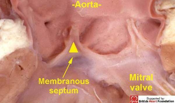
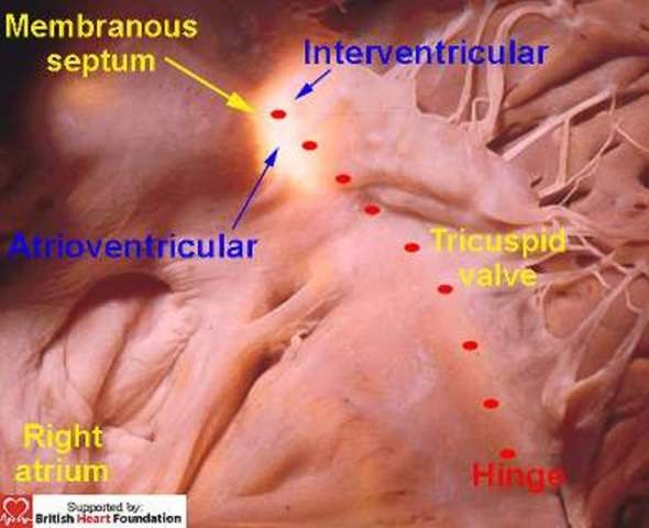
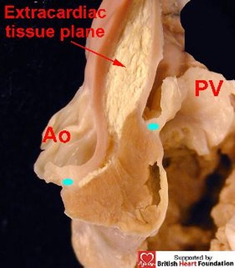
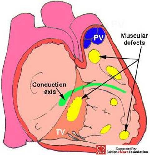
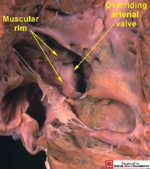
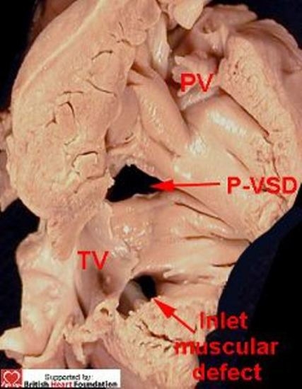
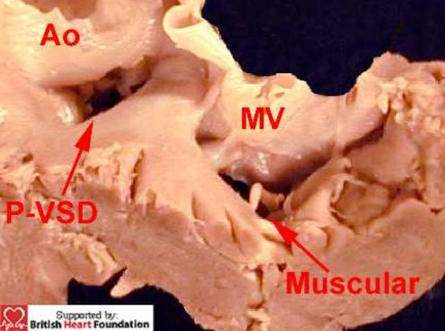
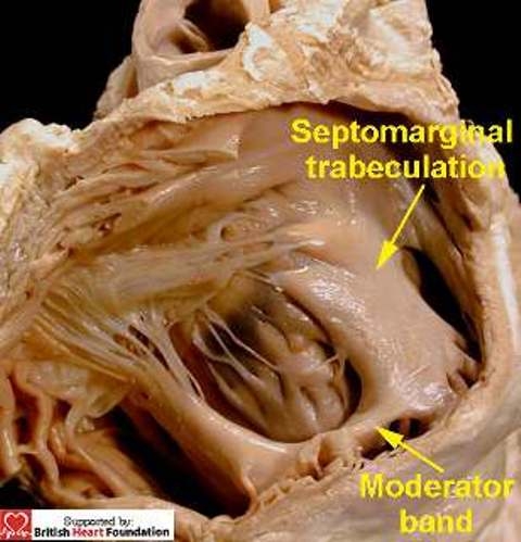
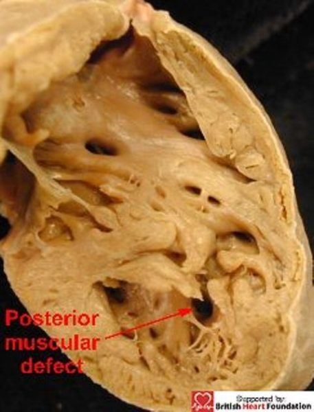
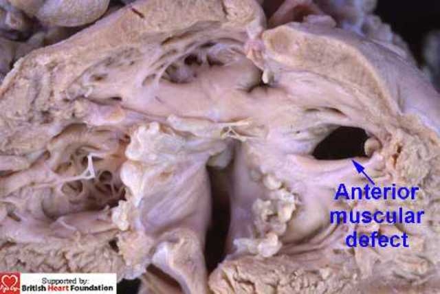
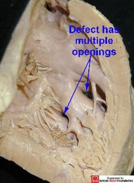
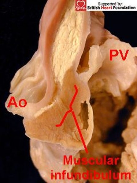
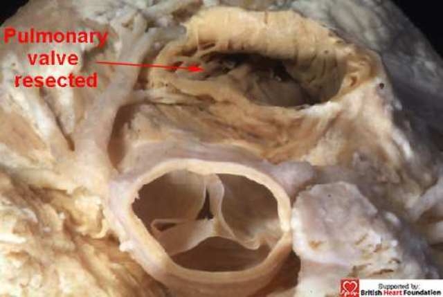
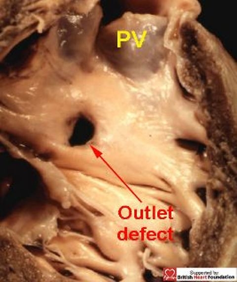
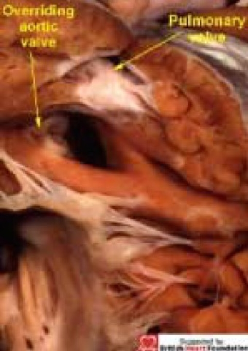
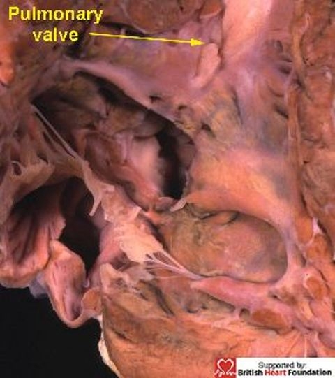
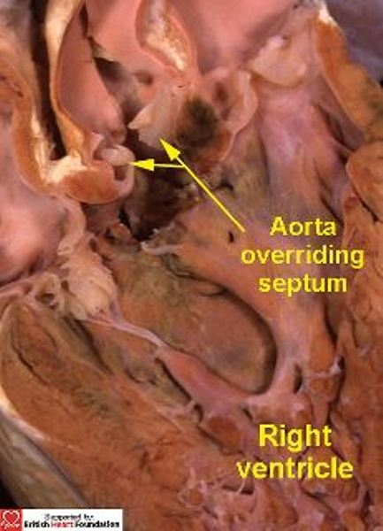
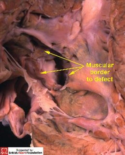
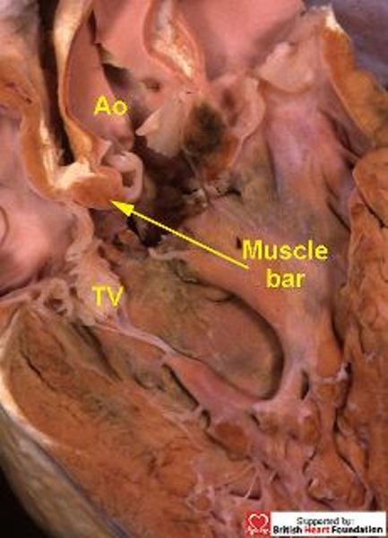
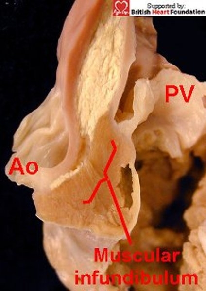
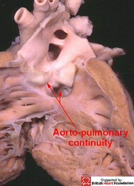
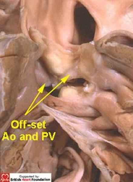
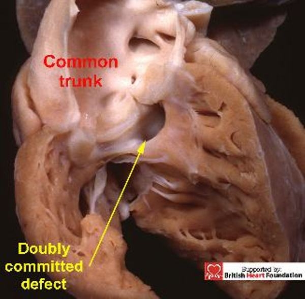
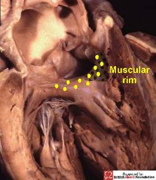
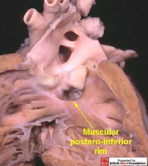
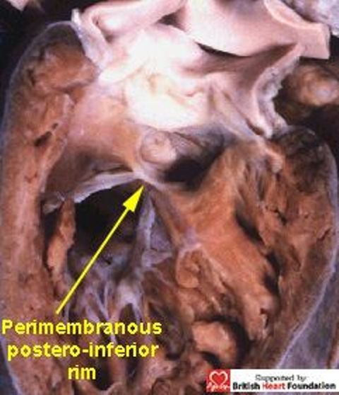
In our previous review of the phenotypic features of ventricular septal defects, we concentrated on the perimembranous variant, showing how its distinguishing feature, as viewed from the right ventricle, was fibrous continuity in its postero-inferior rim between the leaflets of the aortic and tricuspid valves. In this second review, we focus on the morphology of those defects which have exclusively muscular rims when viewed from their right side, and the variant with the phenotypic feature of fibrous continuity between the leaflets of the two arterial valves. As with the defects described as being perimembranous, once they have been characterised, it is the position of the defect relative to the components of the morphologically right ventricle that is the primary determinant of the options and strategies for treatment. Therefore, clarification of the morphology is the key to establishing the related risks for each particular defect.

Keywords: Heart defects; Heart septal defects; Heart ventricle/pathology; Heart ventricles/abnormalities; congenital; ventricular.


Figures

Figure 1
Figure 1
The membranous septum is found at the base of the inter-leaflet triangle which interposes between the right coronary and non-coronary leaflets of the aortic valve
Figure 2
Figure 2
Trans-illumination from the left ventricle shows how the right atrioventricular aspect of the membranous septum is crossed by the hinge of the tricuspid valve. This divides it into an atrial and ventricular components. The conduction axis penetrates through the atrioventricular part of the septum.
Figure 3
Figure 3
A perimembranous septal defect, showing the fibrous postero-inferior rim of the defect penetrated by the conduction axis
Figure 4
Figure 4
A normal heart showing the relationship of the great arteries. The pulmonary trunk is lifted away from the base of the heart by a free-standing muscular infundibulum
Figure 5
Figure 5
The attachment of the hinge points (blue dots) of the aortic (Ao), and the pulmonary valves (PV), are off-set, with an extracardiac tissue plane separating the two arterial trunks. The situation is illustrated here in the setting of tetralogy of Fallot
Figure 6
Figure 6
It is continuity between the leaflet of the aortic and tricuspid valves in the postero-inferior margin which is the hallmark of a perimembranous ventricular septal defect
Figure 7
Figure 7
This diagrammatic representation shows the possible locations of muscular ventricular septal defects, and their relationship to the conduction axis.(TV= tricuspid valve, PV= pulmonary valve)
Figure 8
Figure 8
When viewed from the right ventricle, this ventricular septal defect has a complete muscular rim, despite the overriding of the aortic valve
Figure 9
Figure 9
Both of these ventricular septal defects are closely related to the tricuspid valve. This shows the right ventricular aspect. (PV= pulmonary valve, P-VSD= perimembranous ventricular septal defect)
Figure 10
Figure 10
From the left ventricular aspect, the defect under the mitral valve (MV) is seen to have a muscular rim. This other defect is perimembranous ventricular septal defect (P-VSD) being roofed by fibrous continuity between the aortic valve (Ao) and the atrioventricular valves
Figure 11
Figure 11
This view of the right ventricular aspect of the normal heart shows the septomarginal trabeculation, which gives rise to the moderator band
Figure 12
Figure 12
This muscular ventricular septal defect is posterior to the septomarginal trabeculation
Figure 13
Figure 13
This muscular ventricular septal defect is anterior to the septomarginal trabeculation
Figure 14
Figure 14
This muscular ventricular septal defect, viewed from its left ventricular aspect, is seen to be a solitary defect
Figure 15
Figure 15
From the right ventricular aspect, the defect is crossed by the septomarginal trabeculations giving the appearance of multiple defects
Figure 16
Figure 16
Muscular infundibulum raises the attachment of the pulmonary valve (PV) away from that of the aorta (Ao)
Figure 17
Figure 17
The pulmonary trunk has been resected in this normal heart, along with the free-standing muscular infundibulum. The dissection has not encroached upon the cavity of the left ventricle
Figure 18
Figure 18
An outlet muscular defect, showing its relationship to the pulmonary valve (PV)
Figure 19
Figure 19
A heart with tetralogy of Fallot, with overriding of the aortic valve (Ao), which is connected to both ventricles, and separated from the tricuspid valve (TV) by a muscle bar of the septomarginal trabeculation (SMT)
Figure 20
Figure 20
This specimen has deviation of the of the muscular outlet septum, but with an unobstructed pulmonary outflow tract
Figure 21
Figure 21
Sectioning the heart shown in Fig.20 reveals that the aortic valve overrides the crest of the ventricular septum
Figure 22
Figure 22
Viewed from the right ventricular aspect, the defect, seen in Figs. 20 & 21, has a complete muscular border
Figure 23
Figure 23
The section shows the muscular bar which separates the aorta (Ao) from the tricuspid valve (TV), thus protecting the conduction bundle
Figure 24
Figure 24
In the normal setting, and seen here in tetralogy of Fallot, the arterial valves are separated by a free standing muscular infundibulum, so the hinge-point attachment of the pulmonary valve (PV) is raised away from the aortic root.(Ao)
Figure 25
Figure 25
With doubly committed defects, the arterial valves are at the same level, with fibrous continuity between the leaflets of the aortic and pulmonary valves
Figure 26
Figure 26
Although this defect is doubly committed, the arterial valves are still off-set
Figure 27
Figure 27
By its very nature, the defects seen with common arterial trunk, is doubly committed and juxtaarterial, and is seen here arising overriding the venticular septum
Figure 28
Figure 28
In this heart with common trunk, there is a muscular postero-inferior rim (yellow dots), separating the truncal valve from the tricuspid valve
Figure 29
Figure 29
The postero-inferior rim of the defect is usually muscular in doubly committed defects
Figure 30
Figure 30
In contrast, the doubly committed defect in this heart extends to become perimembranous, with fibrous contunity between the leaflets of the aortic and tricuspid valves, as well as between the aortic and pulmonary valves

References

    1. McCarthy KP, Ho SY, Anderson RH. Categorisation of ventricular septal defects: review of the perimembranous morphology. Images Paediatr Cardiol. 2000;3:21–30. http://www.magnet.mt/health/impaedcard . - PMC - PubMed
    1. Franklin RCG, Anderson RH, Daniëls O, Elliott M, Gewillig MHML, Ghisla R, Krogman ON, Ulmer HE, Stocker FP. The European Paediatric Cardiac Code – The Short List. Cardiol Young. 2000;10(Suppl 1):8–26. - PubMed
    1. Franklin RCG. The European Paediatric Cardiac Code Long List: structure and function. Cardiol Young. 2000;10(Suppl 1):27–146. - PubMed
    1. Jacobs JP, Burke RP, Quintessenza JA, Mavroudis C. Congenital Heart Surgery Nomenclature and Database Project: ventricular septal defect. Ann Thorac Surg. 2000;69(Suppl 4):S25–35. - PubMed
    1. Anderson RH, Ho SY, Becker AE. Anatomy of the human atrioventricular junctions revisited. Anatomical Record. 2000;260:81–91. - PubMed

LinkOut - more resources