. 2001 Apr;3(2):3-18.

Prenatal diagnosis of congenital anomalies

Affiliations

Prenatal diagnosis of congenital anomalies

T Todros et al. Images Paediatr Cardiol. 2001 Apr.

Abstract

Up till the early 1970s, prenatal diagnosis of congenital anomalies was primarily aimed at detecting chromosomal abnormalities by amniocentesis.1. Over the last two decades, prenatal diagnosis has greatly benefited from advances in ultrasound technology and in our ability to detect microscopic and submicroscopic chromosome abnormalities as well as single gene disorders, leading to substantive improvements in detection of such congenital anomalies.2 At present, invasive prenatal diagnosis continues to be the gold standard for pregnancies at increased risk for chromosomal anomaly or other genetic disease, with chorionic villus sampling being the procedure of choice for the first trimester,3 whereas mid-trimester amniocentesis continues to be the most common form of invasive procedure for prenatal diagnosis.4 Still, invasive techniques are restricted to subgroups at risk for anomalies, for whom such time-consuming procedures are believed to be cost-effective, also accounting for procedure-related abortive risks. In the low-risk population prenatal diagnosis generally consists of screening procedures by means of ultrasound and maternal serum biochemistry.

Keywords: congenital; fetal anomalies; fetal echocardiography; heart defects; prenatal diagnosis; ultrasonography.
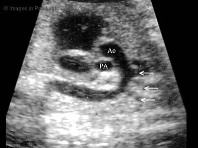
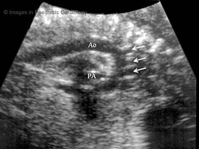
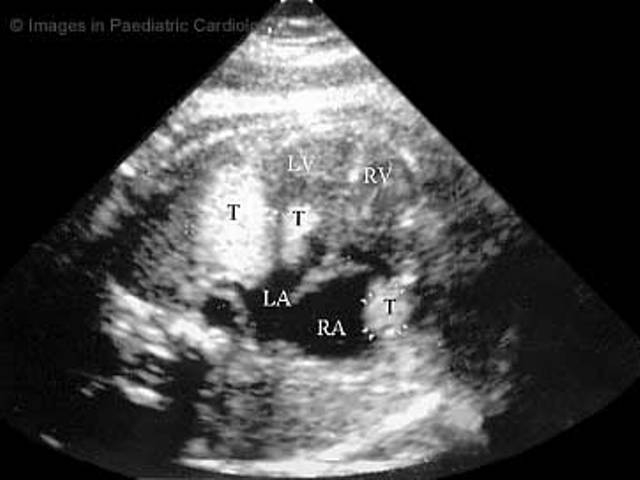
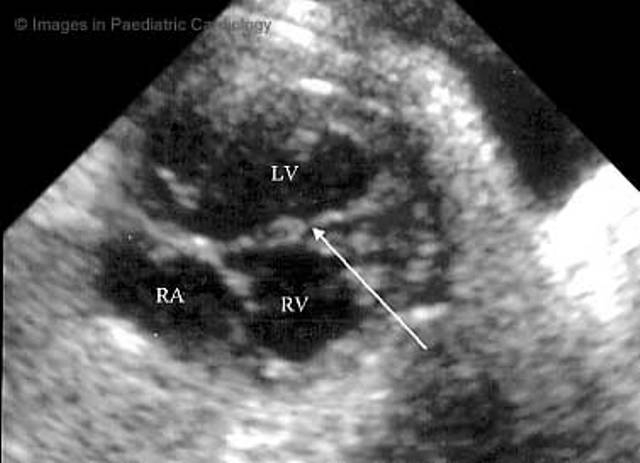

Figures

Fig. 1
Fig. 1
Duodenal atresia at 28 weeks of gestational age.Transverse scan of the abdomen (ST, stomach;D, dilatated duodenal bulb; SP, fetal spine)
Fig. 2
Fig. 2
Umbilical cord at the level of insertion into the fetal abdomen. Hemangioma (arrows); UC, umbilical cord; P, pseudocyst
Fig. 3
Fig. 3
Bilateral polycystic kidney. Transverse section of the abdomen at 28 weeks (K, kidney; S, fetal spine)
Fig 4
Fig 4
Cross section of a fetal thorax with hydrothorax (arrows); L, lung; H, heart
Fig 5
Fig 5
Apical four chamber view of the fetal heart (LV, left ventricle; RV, right ventricle; LA, left atrium; RA, right atrium; MB moderator band; PV, pulmonary veins; D Ao, descending aorta; S, fetal spine)
Fig 6
Fig 6
Short axis of the great vessels (RV, right ventricle; PV, pulmonary valve; PA pulmonary artery; RA, right atrium; Ao, aorta)
Fig. 7
Fig. 7
Long axis Aorta (Ao, aorta; RV, right ventricle; IVS, interventricular septum; LV, left ventricle; LA, left atrium)
Fig. 8
Fig. 8
Ductus arteriosus (D, ductus arteriosus; RV, right ventricle; PA pulmonary artery; D Ao, descending aorta; A Ao, ascending aorta)
Fig. 9
Fig. 9
Right parasagittal scan of fetal trunk demonstrating the inferior and superior venae cavae (IVC, SVC) entering the right atrium (RA)
Fig. 10
Fig. 10
The aortic arch (Ao) as viewed from the anterior toracic wall (PA, pulmonary artery; head and neck vessels are indicated by the small arrows)
Fig. 11
Fig. 11
The aortic arch (Ao) as viewed from the back of the fetus (PA, pulmonary artery; head and neck vessels are indicated by the small arrows)
Fig. 12
Fig. 12
Four chamber apical view in a third trimester fetus with multiple cardiac rhabdomyomas (T, tumor; RV, right ventricle; LV, left ventricle; RA, right atrium; LA, left atrium)
Fig. 13
Fig. 13
Four chamber view reveals a muscular ventricular septal defect (arrow), RV, right ventricle; LV, left ventricle; RA, right atrium; LA, left atrium
Fig. 14-15
Fig. 14-15
Four chamber view and color Doppler of the same fetus reveals bidirectional flow through the ventricular septal defect (arrows)
Fig. 16
Fig. 16
Transposition of great arteries in a 28 weeks fetus (LV, left ventricle; RV, right ventricle; PA, pulmonary artery; Ao, aorta)

Similar articles

Cited by

References

    1. Steele MW, Breg WR. Chromosome analysis of human amniotic fluid cells. Lancet. 1966;1:383–385. - PubMed
    1. Chitayat D, Babul-Hirji R. Genetic counselling in prenatally diagnosed non-chromosomal fetal abnormalities. Curr Opin Obstet Gynecol. 2000;12:77–80. - PubMed
    1. Jenkins TM, Wapner RJ. First trimester prenatal diagnosis: chorionic villus sampling. Semin Perinatol. 1999;23:403–413. - PubMed
    1. Douglas Wilson R. Amniocentesis and chorionic villus sampling. Curr Opin Obstet Gynecol. 2000;12:81–86. - PubMed
    1. In: Fetal Abnormalities. Guidelines for Screeening, Diagnosis and Management. London, UK: RCPCH, RCOG; 1997. Royal College of Obstetricians and Gynaecologists, Royal College of Paediatrics and Child Health. Background evidence supporting the guidelines; p. 5.

LinkOut - more resources