. 2005 Jul;7(3):24-42.

Management of tetralogy of fallot with pulmonary atresia

Affiliations

Management of tetralogy of fallot with pulmonary atresia

Lr Prieto. Images Paediatr Cardiol. 2005 Jul.

Abstract

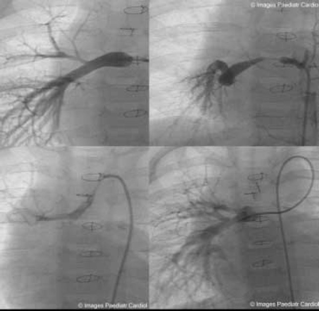
Tetralogy of Fallot with Pulmonary Atresia is an extreme form of tetralogy characterized by absence of flow from the right ventricle to the pulmonary arteries. Pulmonary blood flow is derived from a variety of sources, including native pulmonary artery branches and aorto-pulmonary collaterals with significant variability from patient to patient. Management must therefore be individualized to each patient's anatomy and physiology. Cardiac catheterization plays a crucial diagnostic and therapeutic role in this group of patients. This article is a concise review of the spectrum of anatomic variability seen in this lesion with an emphasis on diagnostic and therapeutic catheterization . It also highlights our staged surgical approach to this lesion and provides data on long-term outcome after complete intracardiac repair.

Keywords: Pulmonary Atresia; Tetralogy of Fallot.


Figures

Figure 1
Figure 1
Angiogram in the left BT shunt in anterior/posterior (A) and lateral (B) projections demonstrates good size right and left pulmonary arteries. This patient had ductal dependent pulmonary circulation and underwent placement of the BT shunt shortly after birth. BT = Blalock Taussig.
Figure 2
Figure 2
Descending aortogram showing two large MAPCA's, one to each lung, and absent native central pulmonary arteries. The patient has a right aortic arch.
Figure 3
Figure 3
A. Descending aortogram in an infant showing multiple MAPCA's to both lungs. B. Selective injection into one of the left-sided MAPCA's fills the native central pulmonary arteries. Note the very stenotic intrapulmonary connection between the MAPCA and the native pulmonary arteries.
Figure 4
Figure 4
A. Injection into a reconstructed right pulmonary artery in a patient who has undergone a right unifocalization. The catheter is inserted via a right modified Blalock Taussig shunt. Note the lack of perfusion to segments of the right upper and right lower lobes in the anterior-posterior projection. B. In the lateral projection the reconstructed right pulmonary artery is seen to perfuse primarily right middle lobe anterior segments.
Figure 5
Figure 5
Selective angiogram of a large MAPCA originating from the left subclavian artery and supplying the left upper lobe in a patient with a right aortic arch.
Figure 6
Figure 6
A. Selective injection in this patient's single coronary artery demonstrating a large MAPCA arising from the proximal left coronary artery branch and perfusing segments of the right upper and right lower lobes. B. Lateral projection of the same injection. MAPCA = major aortopulmonary collateral artery.
Figure 7
Figure 7
Descending aortogram in a ten-year-old patient with TOF/PA who had undergone a central shunt as an infant at an outside institution. Note the significant stenosis in the MAPCA perfusing the left lower lobe, as well as in some of the MAPCA's perfusing the right lung.
Figure 8
Figure 8
Selective angiogram into a large, unrestrictive MAPCA perfusing multiple segments of the left lung as well as some segments of the right lung. This patient is at risk of developing early pulmonary vascular disease if left untreated.
Figure 9
Figure 9
A. Selective angiogram in the patent ductus of a three week old patient who presented with severe cyanosis. The ductus arises from an anomalous left subclavian artery in this patient with a right aortic arch. It supplies the entire left pulmonary artery. At presentation at three weeks of age the ductus was nearly closed and did not respond to prostaglandin. The patient had a stenotic MAPCA to the right lung and saturations in the 40's. B. Following emergent balloon dilation of the ductus, saturations increased into the 70's. A left modified BT shunt was placed ten days later and the ductus was ligated.
Figure 10
Figure 10
A. Selective angiogram in a MAPCA in a three month old shows the severely hypoplastic native pulmonary arteries, which measure at most 1 mm in diameter. 10B: Lateral projection of the same angiogram showing the tiny central pulmonary arteries extending anteriorly. 10C: Pulmonary artery angiogram in the same patient at 18 months of age after a Melbourne shunt at three months of age and left unifocalization with placement of a 4 mm left BT shunt at 11 months of age. 10D: Lateral projection of the same angiogram performed via the left BT shunt. Note the dramatic increase in size of the central pulmonary arteries. The catheter course is from the left BT shunt into the central pulmonary arteries, and then through the Melbourne shunt into the ascending aorta. The catheter is pulled back towards the pulmonary arteries during the injection. The patient underwent complete repair at 2 ½ years of age. 10E, F. Pulmonary angiogram in the same patient at 3 ½ years of age, one year after complete repair. She underwent dilation of left middle and left lower lobe branches with RV pressure decreasing from ¾ systemic to 2/3 systemic. BT = Blalock-Taussig, MAPCA = major aortopulmonary collateral artery, RV = right ventricle.
Figure 11
Figure 11
Wedge angiography in the left upper pulmonary vein reveals absence of a central left pulmonary artery. Note the intraparenchymal left pulmonary artery branches with no filling of any central vessel.
Figure 12
Figure 12
A,B. Selective angiogram in the left pulmonary artery (A) and right pulmonary artery (B) accessed via the Melbourne shunt. Note that a wire has been inserted into the distal right pulmonary artery in order to maintain catheter position in the proximal right pulmonary artery.
Figure 13
Figure 13
A-D. Right unifocalization. A. Selective injection in the right pulmonary artery via a left BT shunt demonstrates a good size branch to the right lower lobe and very small branch to the right upper lobe. B. Selective injection in a MAPCA to the right lower lobe in the same patient. Note the presence of severe mid segment stenosis. C. Additional MAPCA from the underside of the aortic arch to the right upper lobe with evidence of proximal stenosis. D. Selective injection in the right pulmonary artery following unifocalization of the two right-sided MAPCA's showing significantly improved flow to the right upper lobe, as well as increased perfusion to segments of the right lower lobe. There are no significant peripheral stenoses visible. Click to view angiogram. BT = Blalock-Taussig, MAPCA = major aortopulmonary collateral artery.
Figure 14
Figure 14
Angiography in the right pulmonary artery in anterior/posterior (A) and lateral (B) projections in a patient with absent native central pulmonary arteries following bilateral unifocalizations. A neo right pulmonary artery has been constructed into which the BT shunt is inserted. Note extension of the neo right pulmonary artery anteriorly in front of the airway in the lateral projection. The reconstructed right pulmonary artery has been brought into the mediastinum and tacked to the ascending aorta. (C), (D). Angiography in the left pulmonary artery in the same patient. A left BT shunt was inserted into the unifocalized LPA after anastomosing a large MAPCA to the LPA remnant within the lung. The patient underwent complete repair at 16 months of age using a pericardial roll to join the reconstructed right and left pulmonary arteries and a Hancock conduit was placed from the right ventricle to the pericardial roll. 14E,F. Angiogram in AP/cranial (E) and lateral projections (F) in the RV outflow tract 1 ½ years after complete repair. RV pressure was 2/3 systemic. 14G 14H 14I 14J . Peripheral stenoses in the right upper lobe branch and in the distal main RPA (G – anterior/posterior, H – lateral) were dilated with RV pressure decreasing to 55% systemic post dilation (I – anterior/posterior, J – lateral). AP = anterior/posterior, BT = Blalock-Taussig, LPA = left pulmonary artery, MAPCA = major aortopulmonary collateral artery, RPA = right pulmonary artery, RV = right ventricle. Click to view angiogram.
Figure 15
Figure 15
A. Right pulmonary artery angiogram demonstrating significant long segment stenosis. The stenosis extends fairly distally into the hilum, best appreciated in the lateral projection (B). Initial angiography was performed with a catheter advanced retrograde through the central shunt. C, D. Following balloon angioplasty, there is marked improvement in the stenosis. Note the catheter is now accessing the shunt in a prograde fashion from the right ventricle to the ascending aorta and through the central shunt. By accessing the pulmonary artery from a venous approach to perform balloon angioplasty, it was possible to avoid placement of a large sheath in the femoral artery.
Fig. 16
Fig. 16
Right pulmonary artery angiogram before (A) and after (B) balloon angioplasty in a patient two months after complete repair. The left pulmonary artery was also dilated. Right ventricular pressure decreased from 80% systemic to 60% systemic.

References

    1. Haworth SG, Macartney FJ. Growth and development of pulmonary circulation in pulmonary atresia with ventricular septal defect and major aorto-pulmonary collateral arteries. British Heart Journal. 1980;44:14–24. - PMC - PubMed
    1. Faller K, Haworth SG, Taylor JFN, Macartney FJ. Duplicate sources of pulmonary blood supply in pulmonary atresia with ventricular septal defect. British Heart Journal. 1981;46:263–268. - PMC - PubMed
    1. Thiene G, Frescura C, Bini RM, Valente M, Gallucci V. Histology of pulmonary arterial supply in pulmonary atresia with ventricular septal defect. Circulation. 1979;60:1066–1074. - PubMed
    1. Haworth SG. Collateral arteries in pulmonary atresia with ventricular septal defect. British Heart Journal. 1980;44:5. - PMC - PubMed
    1. Iyer KS, Mee RB. Staged repair of pulmonary atresia with ventricular septal defect and major systemic to pulmonary artery collaterals. Ann Thorac Surg. 1991;51:65–72. - PubMed

LinkOut - more resources