

A brief history of cardiac pacing
Images Paediatr Cardiol.
2006 Apr.
Abstract
This article is the first of three articles that will deal with pacing. The history and background leading to pacemakers as we know them is briefly discussed.
Figures
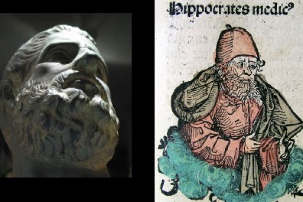
Hippocrates (460 - 375 BC)
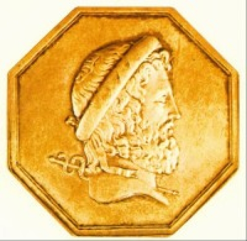
Aristotle (384 - 322 BC)
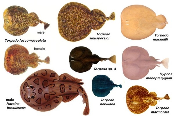
Electric Rays
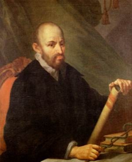
Geronimo Mercuriale (1530 - 1606)
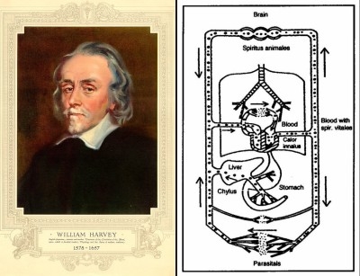
William Harvey and the circulation
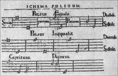
A pulse diagram by Valentini
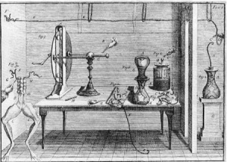
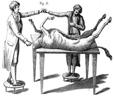
Stimulation device (1788)
Luigi Galvani (1737 - 1798)
Galvani's electrostatic nerve stimulator
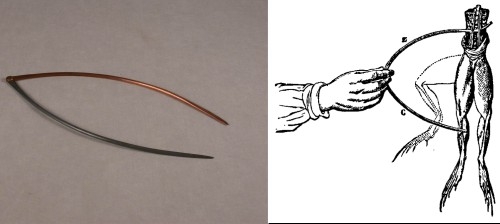
Galvani's forceps
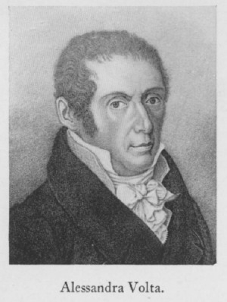
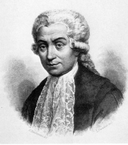
Alessandro Volta (1745 - 1827)
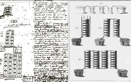
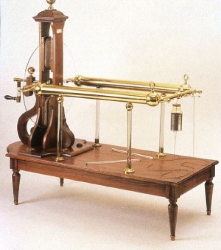
The Volta Pile
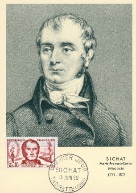
Marie Francois Xavier Bichat (1701 - 1802)
Aldini (1762 - 1834)
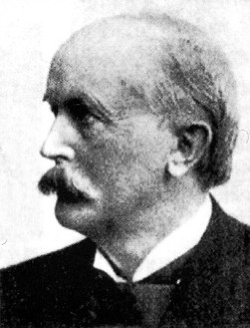
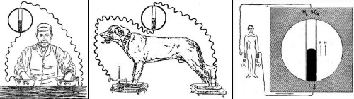
Hugo Von Ziemssen (1829 - 1902)
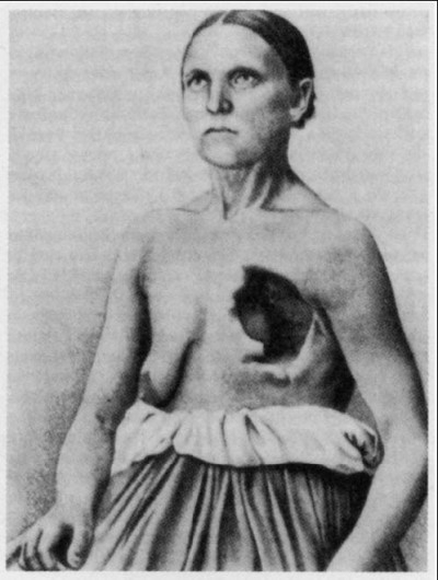
Catharina Serafin
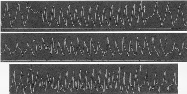
Catharina's cardiac activity
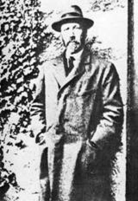
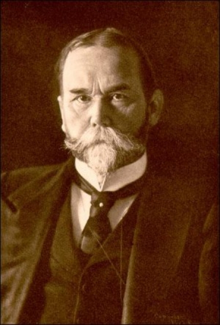
John Mac William
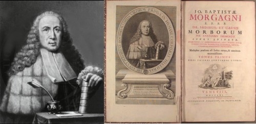
Giovanni Battista Morgagni (1682 - 1771)
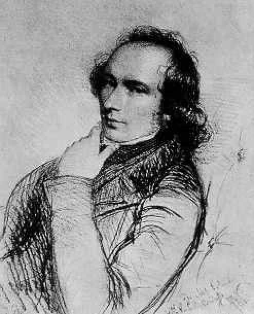
William Stokes (1804 - 1878)
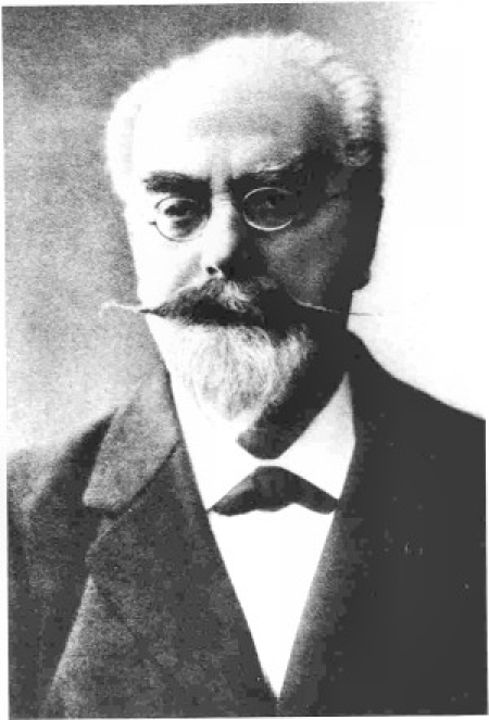
Karel Frederik Wenckebach
John Hay
Augustus Desire Waller
Lippmann

Lippmann's electrometer
Waller's recording setup
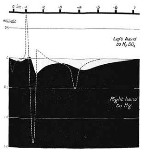
First ECG
Early Waller ECG's and apparatus
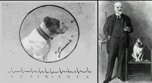
Jimmie with its proud owner
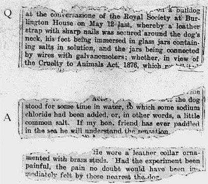
From the records of the House of Commons
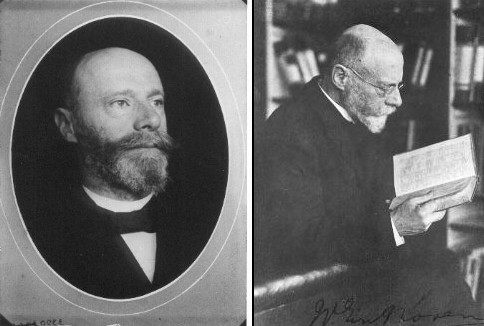
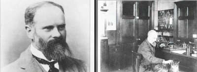
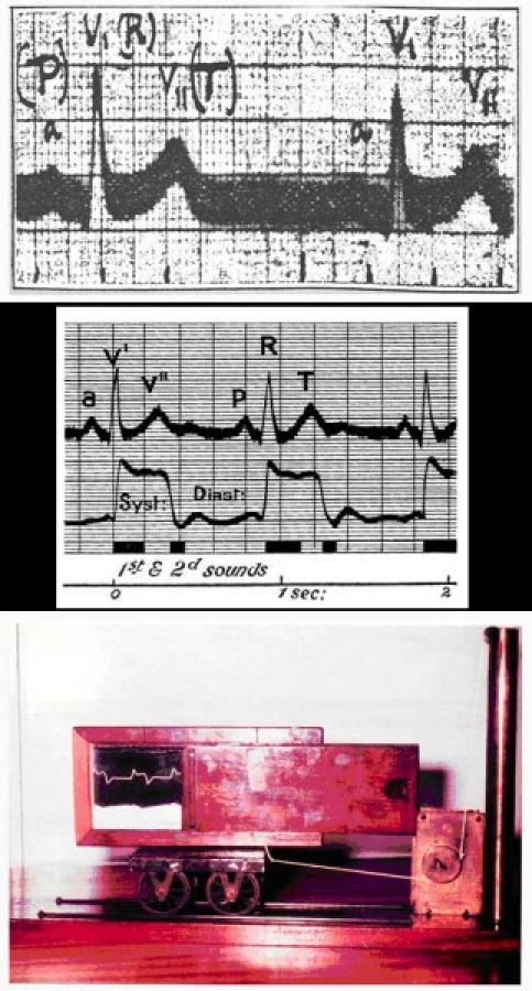
Willem Einthoven (1860 – 1927)
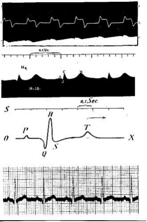
Early Einthoven ECG's
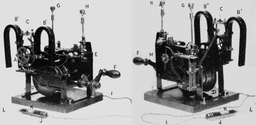
The String Galvanometer
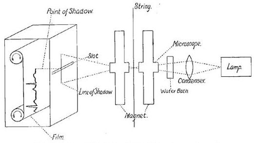
Einthoven's recording schema
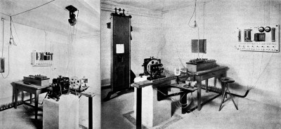
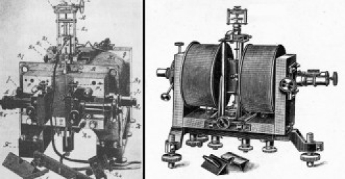
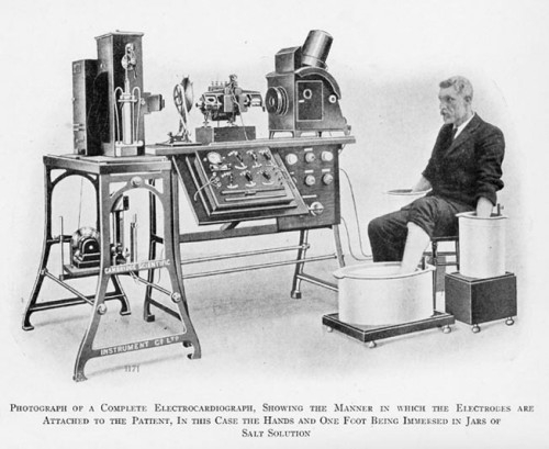
Einthoven's lab setup with the string galvanometer (two views)
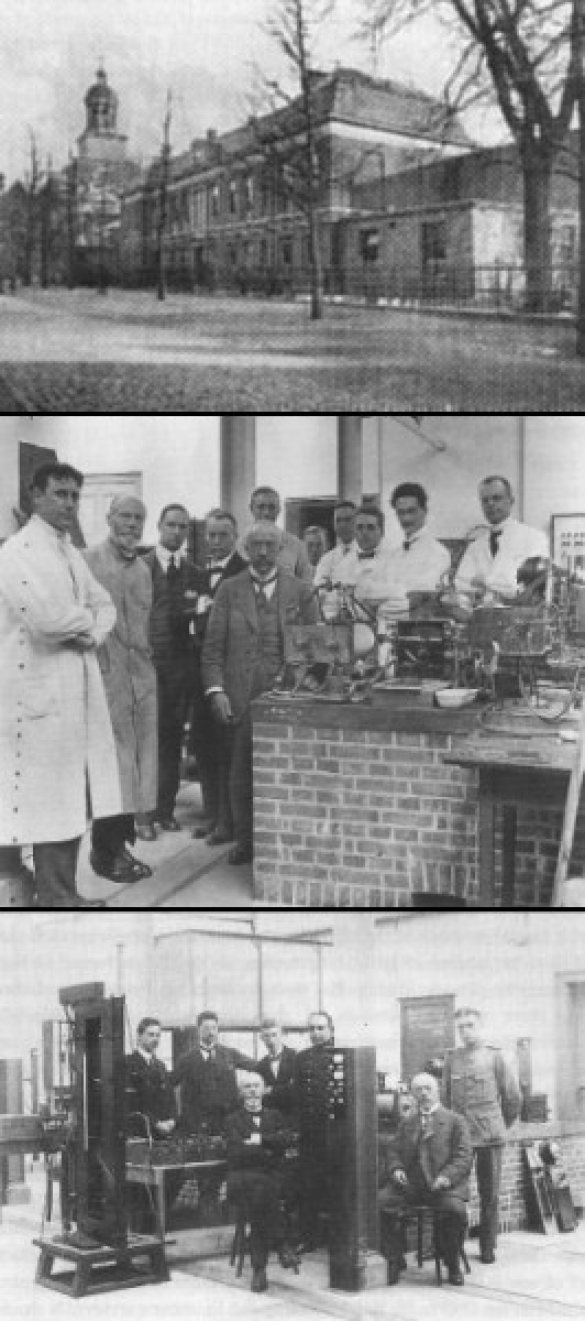
Einthoven's lab and colleagues
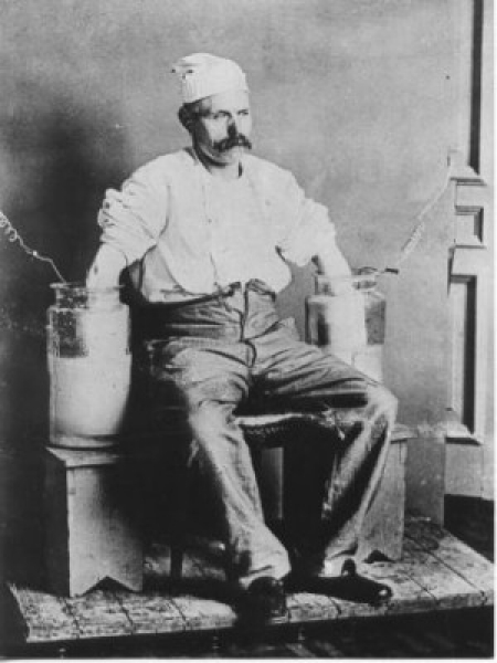
Early ECG recording technique
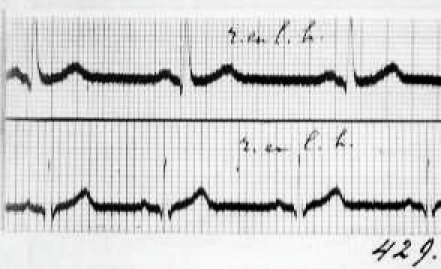
Early Einthoven ECG
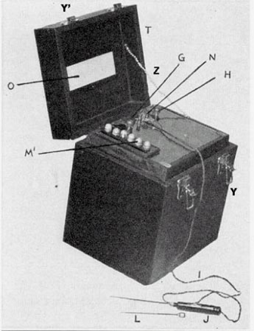
Early ECG machine
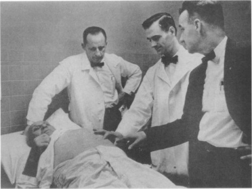
Albert Hyman's “artificial pacemaker”: the two photos
Hyman's device in a box
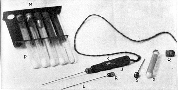
Hyman's electrodes
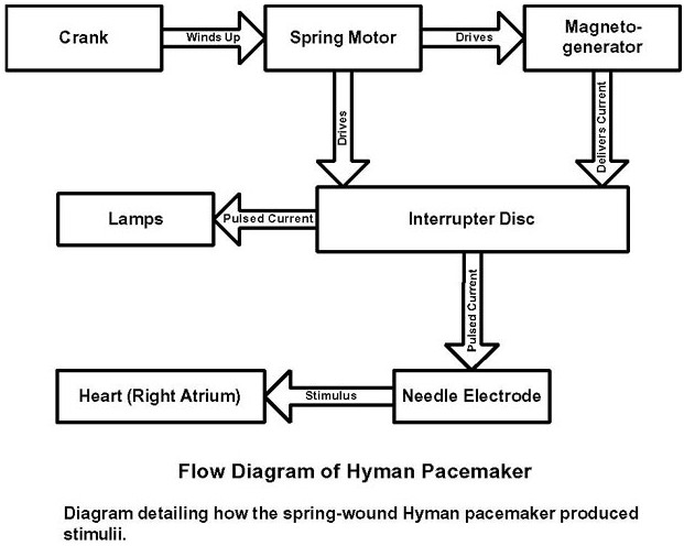
Flow diagram of Hyman's “artificial pacemaker”
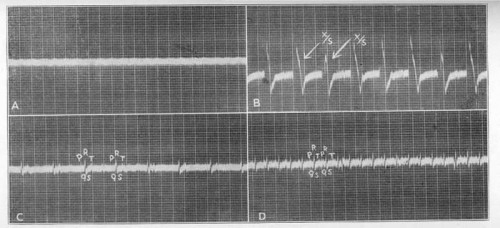
Hyman's recordings
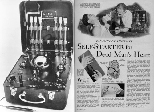
The Hymanotor (Adlanco)
Wilfred Bigelow
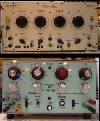
The Grass stimulator
John Hopps
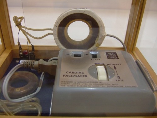
Early external electronic pacemakers
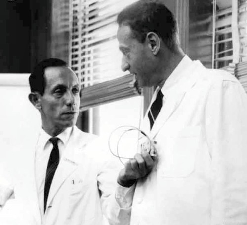
Paul Zoll and a colleague
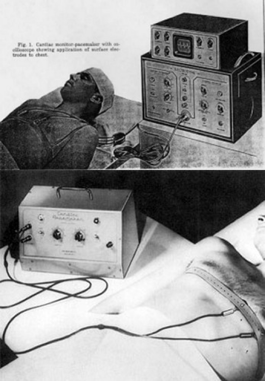
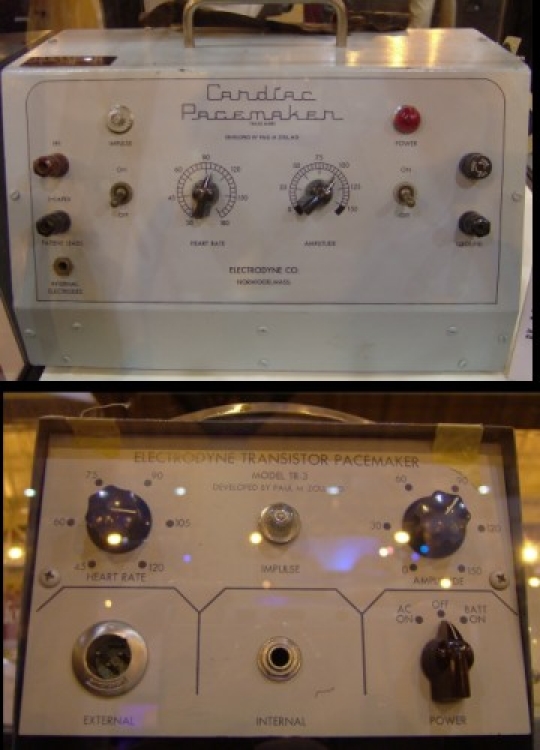
Zoll's external pacemaker
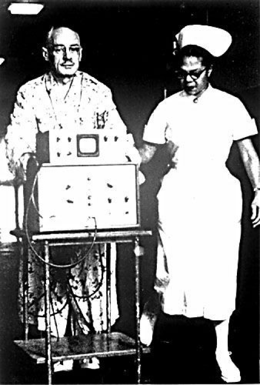
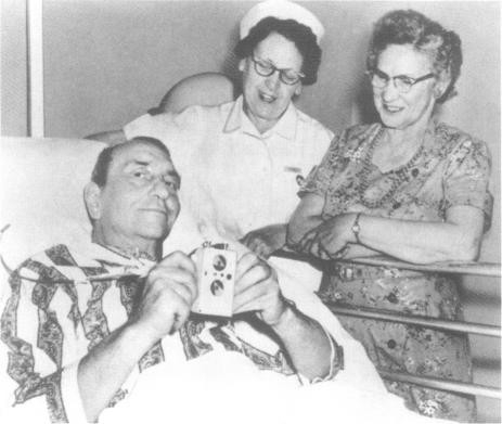
The PM-65: historic 1958 photo (patient was using the first catheter electrode)
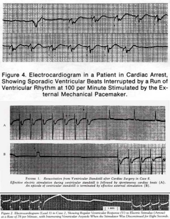
Early Zoll paced ECG tracings
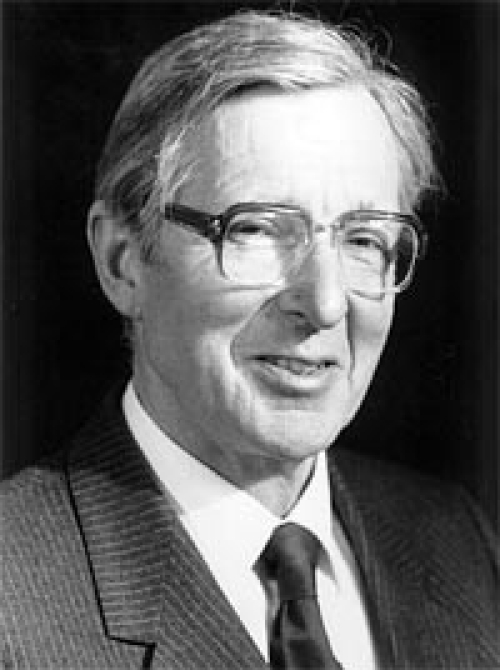
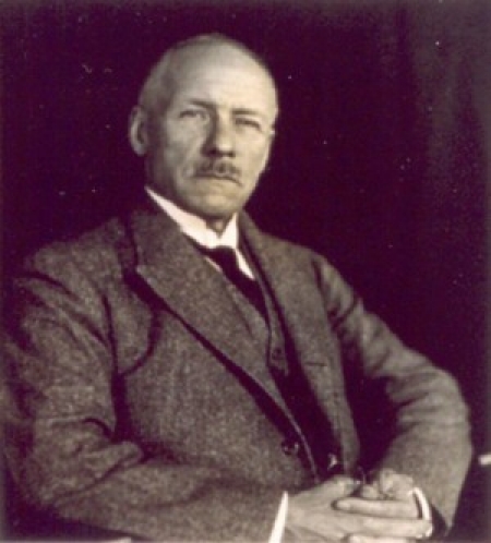
Aubrey Leatham
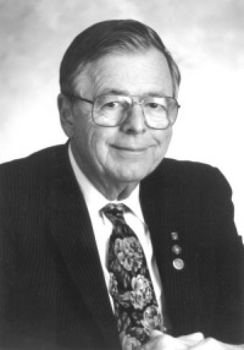
Earl Bakken
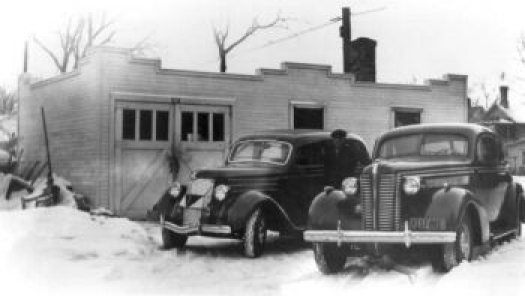
Medtronic's first building
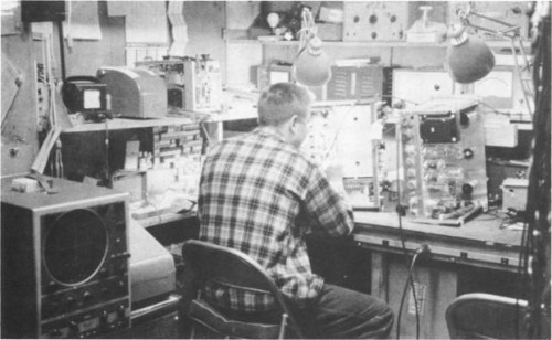
Bakken's first lab

Walton Lillehei
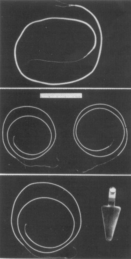
Myocardial pacing wire and indifferent electrode
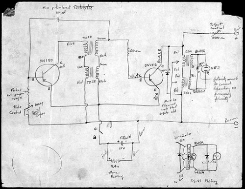
Bakken's circuit
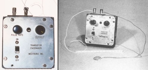
Bakken's pacemaker with leads
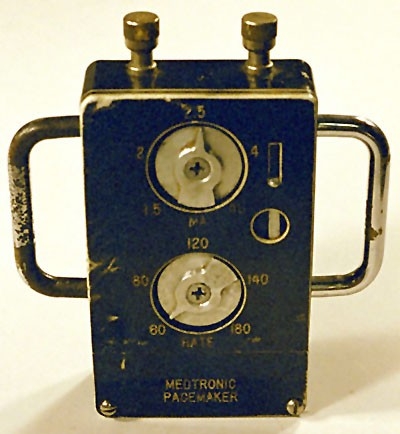
One of the “first ten”
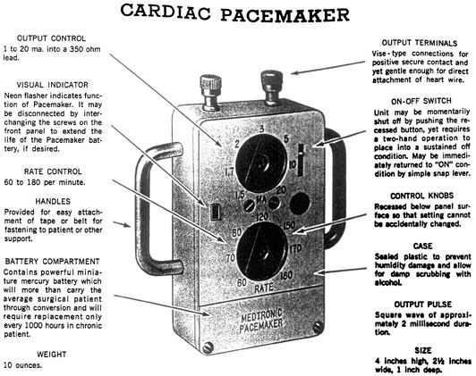
Product literature of the “first ten”
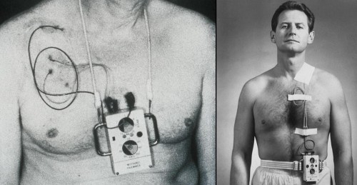
Wearable devices on patients (1958)

Lillehei with a child being paced
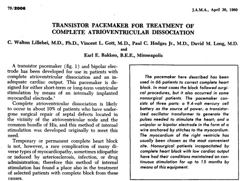
Lillehei's paper
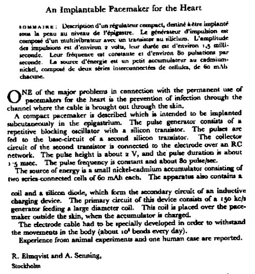
Senning's and Elmqvist's paper
Ake Senning
Rune Elmqvist
Arne Larsson
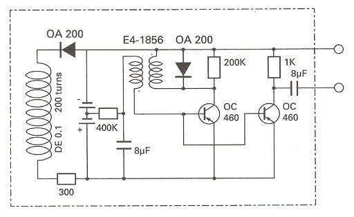
Elmqvist's circuit
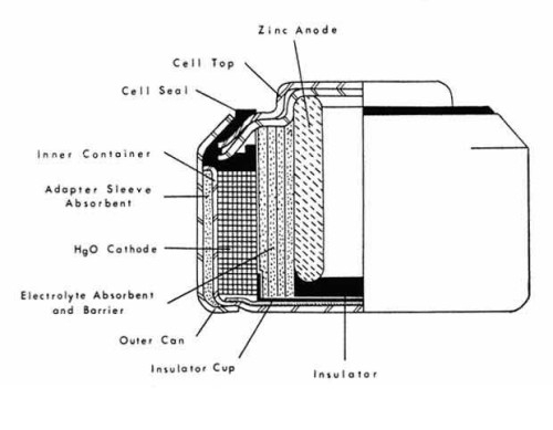
Mercury Cell
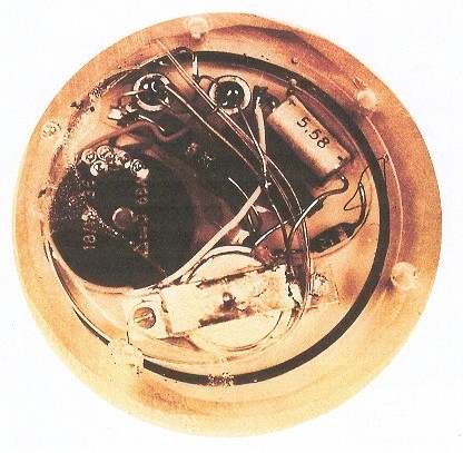
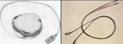
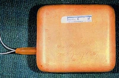
First implanted pacemaker
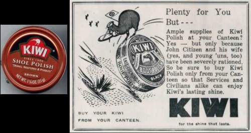
Kiwi Shoe Polish
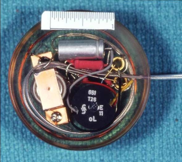
Modern replica of Elmqvist's pacemaker
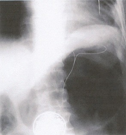
X-ray of Larsson showing pacemaker and leads
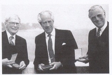
Elmqvist, Senning and Larsson (left to right)
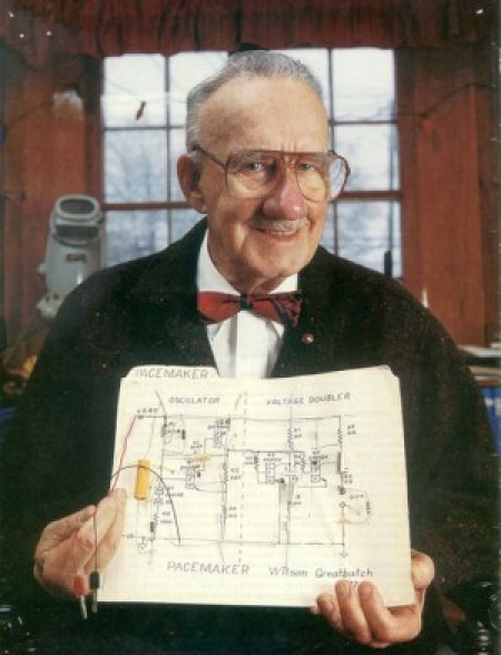
Wilson Greatbatch and his circuit
The “Bow Tie Team”
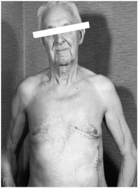
Patient with Greatbatch's pacemaker
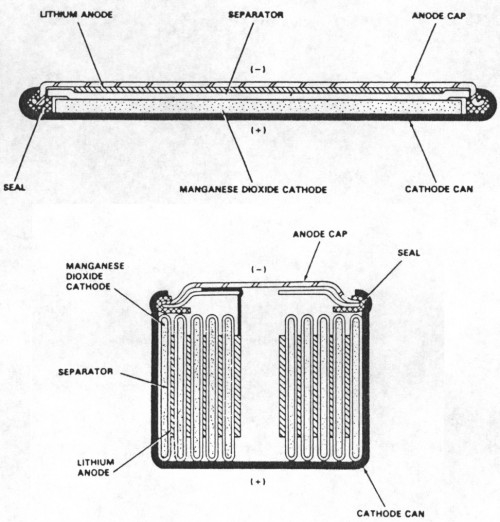
Lithium-Iodine Cell
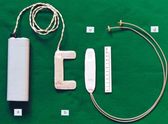
Inductively-coupled pacing
External coil and device for inductive device
Warren Mauston, the first recipient of the Hunter-Roth electrode
Hunter-Roth electrodes
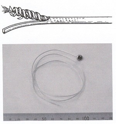
Elema lead
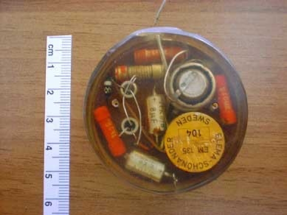
The Elema 135
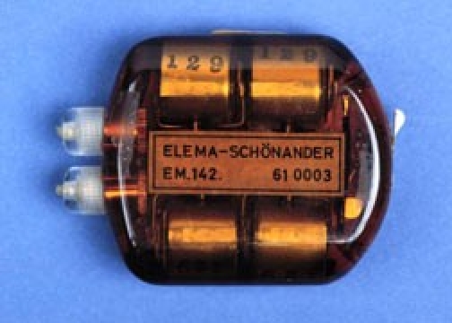
The Elema 142 (with non-rechargable cells)
Zoll device
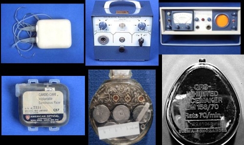
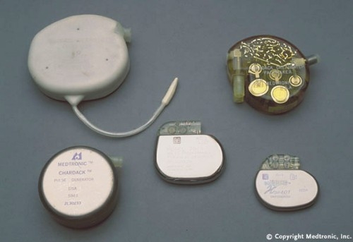
Devices of the 60's
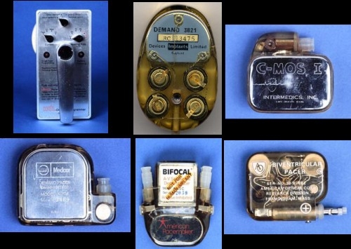
Devices of the 70's
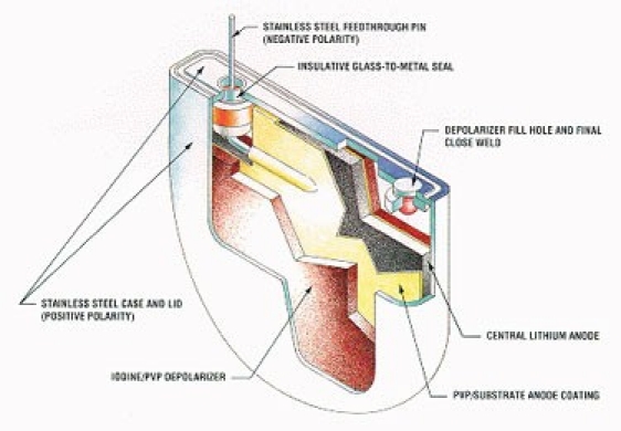
Modern lithium pacemaker cell
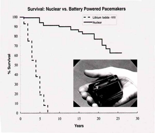
Nuclear pacemakers with projected longevity
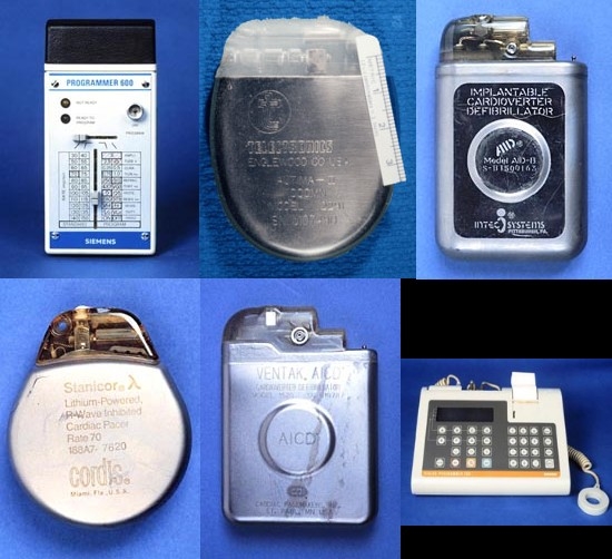
Devices of the 80's
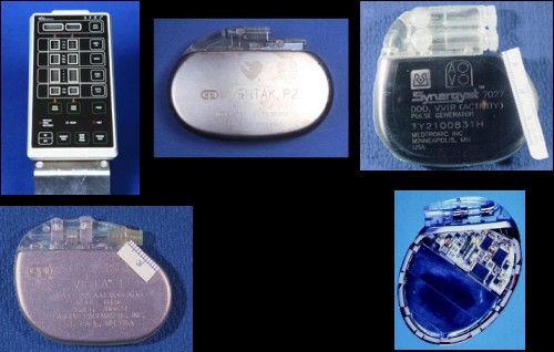
Devices of the 90's
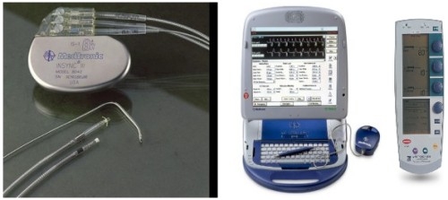
Contemporary devices
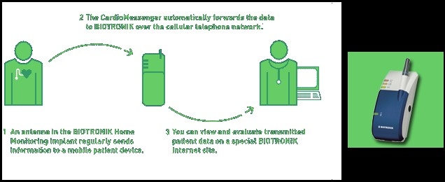
Home monitoring via the World Wide Web
History of pacing
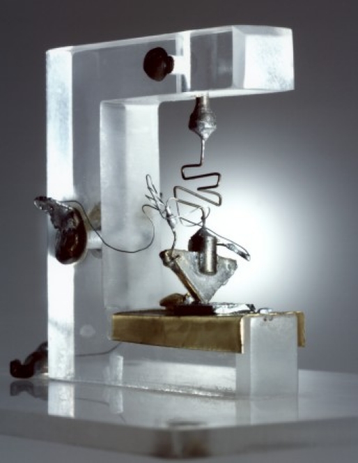
The “star” of pacing: the first transistor
LinkOut - more resources
Full Text Sources
Other Literature Sources