. 2011 Dec 21;2(12):730-742.
doi: 10.1021/cn200090z.

The Discovery and Characterization of ML218: A Novel, Centrally Active T-Type Calcium Channel Inhibitor with Robust Effects in STN Neurons and in a Rodent Model of Parkinson's Disease

Affiliations

The Discovery and Characterization of ML218: A Novel, Centrally Active T-Type Calcium Channel Inhibitor with Robust Effects in STN Neurons and in a Rodent Model of Parkinson's Disease

Zixiu Xiang et al. ACS Chem Neurosci. .

Abstract

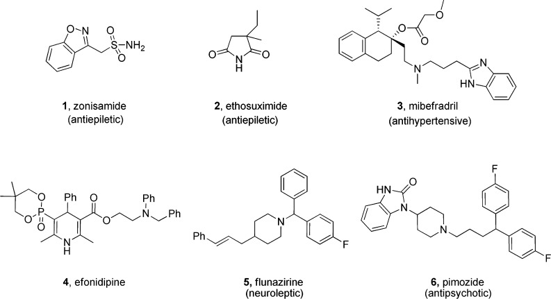
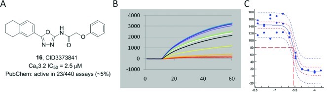
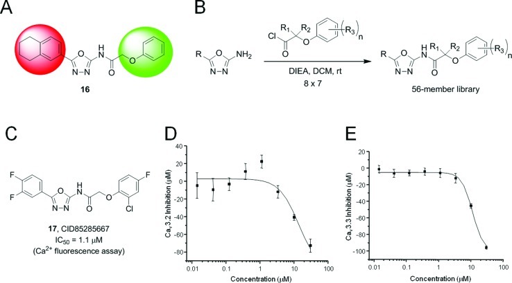
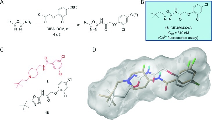
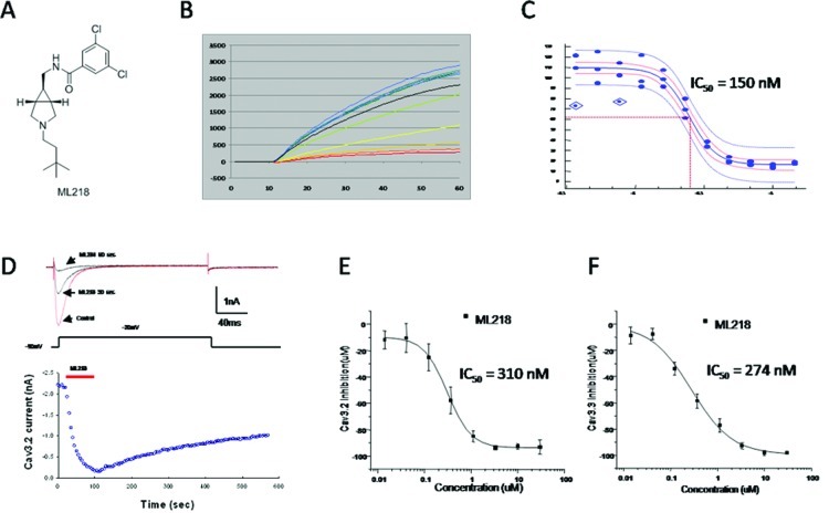
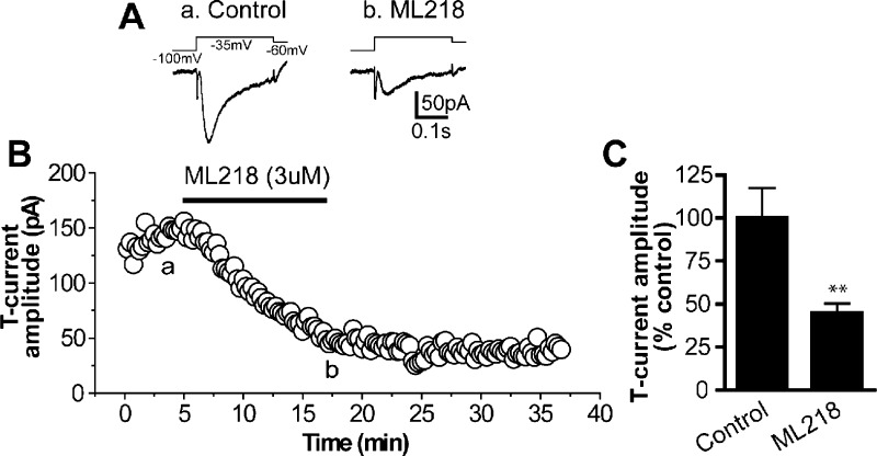
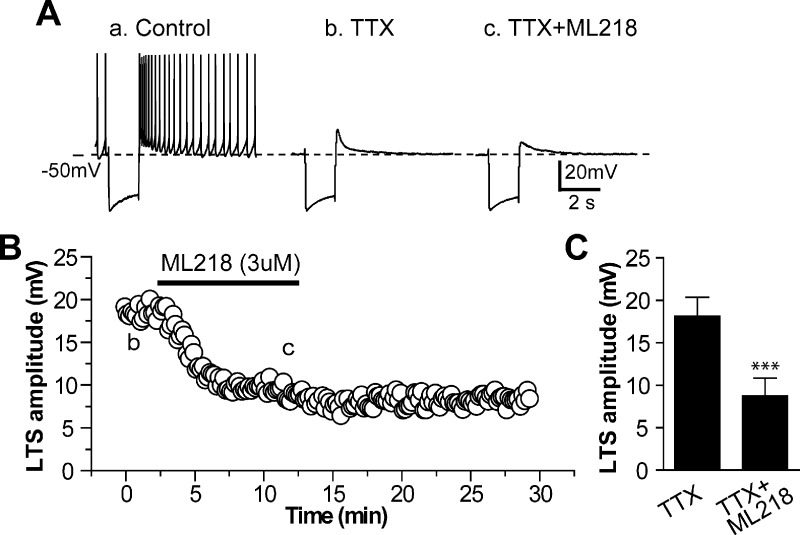
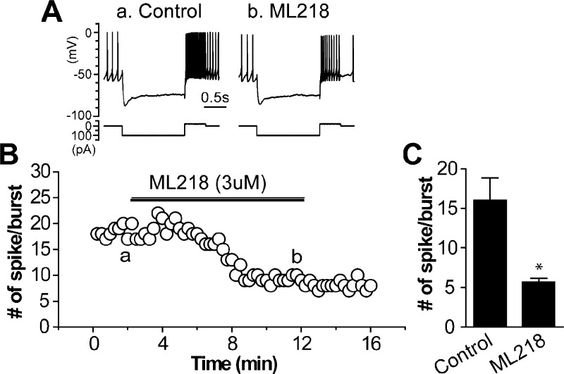
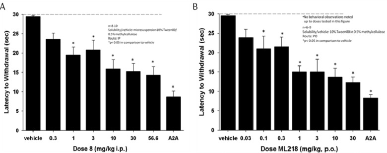
T-type Ca(2+) channel inhibitors hold tremendous therapeutic potential for the treatment of pain, epilepsy, sleep disorders, essential tremor and other neurological disorders; however, a lack of truly selective tools has hindered basic research, and selective tools from the pharmaceutical industry are potentially burdened with intellectual property (IP) constraints. Thus, an MLPCN high-throughput screen (HTS) was conducted to identify novel T-type Ca(2+) channel inhibitors free from IP constraints, and freely available through the MLPCN, for use by the biomedical community to study T-type Ca(2+) channels. While the HTS provided numerous hits, these compounds could not be optimized to the required level of potency to be appropriate tool compounds. Therefore, a scaffold hopping approach, guided by SurflexSim, ultimately afforded ML218 (CID 45115620) a selective T-Type Ca(2+) (Ca(v)3.1, Ca(v)3.2, Ca(v)3.3) inhibitor (Ca(v)3.2, IC(50) = 150 nM in Ca(2+) flux; Ca(v)3.2 IC(50) = 310 nM and Ca(v)3.3 IC(50) = 270 nM, respectively in patch clamp electrophysiology) with good DMPK properties, acceptable in vivo rat PK and excellent brain levels. Electrophysiology studies in subthalamic nucleus (STN) neurons demonstrated robust effects of ML218 on the inhibition of T-Type calcium current, inhibition of low threshold spike and rebound burst activity. Based on the basal ganglia circuitry in Parkinson's disease (PD), the effects of ML218 in STN neurons suggest a therapeutic role for T-type Ca(2+) channel inhibitors, and ML218 was found to be orally efficacious in haloperidol-induced catalepsy, a preclinical PD model, with comparable efficacy to an A(2A) antagonist, a clinically validated PD target. ML218 proves to be a powerful new probe to study T-Type Ca(2+) function in vitro and in vivo, and freely available.


Figures

Figure 1
Figure 1
First generation T-type Ca2+ inhibitors derived from marketed antiepileptic 1 and 2, antihypertensives 3 and 4, neuroleptic 5, and antipsychotic 6 drugs.
Figure 2
Figure 2
Second generation T-type Ca2+ inhibitors 715 derived from optimization of HTS hits.
Figure 3
Figure 3
Confirmed HTS hit 16. (A) Structure of 16, CID3373841. (B) Raw fluorescence-based HTS assay traces. (C) CRC from fluorescence-based assay (IC50 = 2.5 μM).
Figure 4
Figure 4
(A) Chemical optimization plan for 16, a two-dimensional library. (B) 8 × 7 member library design that generated 56 analogues of 16. (C) Structure of 17 (CID85285667), the most potent analogue with an IC50 of 1.1 μM in the Ca2+ fluorescence assay. (D) Cav3.2 IonWorks Quattro (patch EP) CRC IC50 = 13.5 μM. (E) Cav3.3 IonWorks Quattro (patch EP) CRC IC50 = 11.5 μM.
Figure 5
Figure 5
Second generation library of analogues of 17. (A) 4 × 2 library of branched alkyl analogues. (B) Structure and activity of most potent analogue, 18 (CID46943243). (C) Manual overlay of 18 with Merck’s 8. (D) SurflexSim flexible alignment of 18 (gray) with Merck’s 8 (tan), affording reasonable overlap except for amide HBD/HBA.
Figure 6
Figure 6
Initial “scaffold hopping” library. 24-membered library of 3-amino piperidine “scaffold hopping” libraries leading to 23 (CID45115608), displaying enantiospecific T-type Ca2+ channel inhibition.
Scheme 1
Scheme 1. Synthesis of [3.1.0] Analogues 26
Figure 7
Figure 7
Second generation library of analogues of 18 focusing on a [3.1.0] core. (A) Structure and activities of [3.1.0] analogues 26ah. (B) Manual overlay of 18 and 26b with Merck’s 8. (C) SurflexSim flexible alignment of 26b (gray) with Merck’s 8 (tan).
Figure 8
Figure 8
Dose–response curves for ML218 (26b). (A) Structure of ML218 (26b). (B) Raw fluorescence-based HTS assay traces. (C) CRC from fluorescence-based assay (IC50 = 150 nM). (D) Inhibition time-course of ML218. Ba2+ currents were elicited by pulse depolarized to −20 mV from a holding potential of −90 mV (200 ms, 0.2 Hz). The bath application of ML218 caused rapid inhibition and slow partially recovery after washout (n = 5). (E) Cav3.2 IonWorks Quattro (patch EP) CRC IC50 = 310 ± 15 nM. (F) Cav3.3 IonWorks Quattro (patch EP) CRC IC50 = 274 ± 53 nM.
Figure 9
Figure 9
ML218 inhibits T-type calcium currents in STN neurons. (A) Averaged traces of T-currents (lower) elicited by a voltage clamp protocol (upper) in control (a) and after application of 3uM ML218 (b) in a voltage clamp experiment. (B) Time course of T-current amplitude before and after application of 3 uM ML218 from the same cell as in (A). (a) and (b) indicate the time points at which averaged traces were taken. (C) Bar graph summarizes group data showing ML218 inhibits T-currents in STN neurons (45.1 ± 5.1% of the control value, n = 7, **p < 0.005). Note that the inhibition persists 20 min after washout of ML218.
Figure 10
Figure 10
ML218 inhibits low threshold spike (LTS) in STN neurons. (A) Representative voltage responses to intracellular injection of a hyperpolarizing current pulse (−160 pA) in control (a), in the presence of 0.5 uM TTX (b), and combination of 0.5 uM TTX and 3 uM ML218 (c) from a current clamp experiment, showing the typical rebound burst firing following the termination of hyperpolarizing current pulse in control (a), pharmacologically isolated LTS in the presence of TTX (b), and inhibition of LTS by ML218 (c). (B) Time course of the effect of ML218 on amplitude of LTS obtained from the same STN neuron as in (A). (b) and (c) indicate the time points at which sample traces were taken. (C) Bar graph summarizes the group data showing ML218 inhibits the amplitude of LTS (8.7 ± 2.1 mV with ML218, compared to 18.1 ± 2.2 mV in control, n = 5, ***p < 0.0001).
Figure 11
Figure 11
ML218 reduces rebound burst activity in STN neurons. (A) Representative voltage responses (upper) to intracellular injection of hyperpolarizing (−100 pA) followed by depolarizing (+20 pA) current pulses (lower) in control and after application of 3uM ML218 from a current clamp experiment. (B) Time course of the number of rebound spikes during the depolarizing current pulse before and after application of 3 uM ML218 from the same cell as in (A). (a) and (b) indicate the time points at which sample traces were taken. (B) Bar graph summarizes the group data showing ML218 reduces the number of rebound spikes in STN neurons (5.7 ± 0.5 spikes/burst with ML218, compared to 16.0 ± 2.8 spikes/burst in control, n = 6, *p < 0.05).
Figure 12
Figure 12
T-Type Ca2+ channel antagonist 8 and ML218 produce a dose-dependent reversal of haloperidol (0.75 mg/kg, i.p.)-induced catalepsy in rats. (A) For comparison, the effects of increasing dose of 8 (i.p.) were compared to a top dose (56.6 mg/kg p.o.) of a previously published A2A antagonist from Neurocrine. (B) Comparison of the effects of increasing dose of ML218 (p.o.) to the Neurocrine A2A antagonist at 56.6 mg/kg (p.o.). Catalepsy was measured as the latency to withdraw the forepaws from a horizontal bar with a cutoff of 30 s. Bar graphs represent the means ± SEM of either 6–10 rats/treatment group (*p < 0.05 versus the vehicle control group by Dunnett’s test).

References

    1. Petegem F. V.; Minor D. L. Jr. (2006) The structural biology of voltage-gated calcium channel function and regulation. Biochem. Soc. Trans. 34, 887–893. - PMC - PubMed
    1. Huguenard J. R. (1998) Low-voltage-activated (T-type) calcium-channel genes identified. Trends Neurosci. 21, 451–452. - PubMed
    1. Ertel E. A.; Campbell K. P.; Harpold M. M.; Hofmann F.; Mori Y.; Perez-Reyes E.; Schwartz A.; Snutch T. P.; Tanabe T.; Birnbaumer L.; Tsien R. W.; Catterall W. A. (2005) Nomenclature of voltage-gated calcium channels. Neuron 25, 533–535. - PubMed
    1. Catterall W. A.; Perez-Reyes E.; Snutch T. P.; Striessing J. (2005) International Union of Pharmacology. XLVIII. Nomenclature and structure-function of voltage-gated calcium channels. Pharmacol. Rev. 57, 411–425. - PubMed
    1. Catterall W. A. (2000) Structure and regulation of voltage-gated Ca2+ channels. Annu. Rev. Cell Dev. Biol. 16, 521–555. - PubMed

LinkOut - more resources