

The sub-pial resection technique for intrinsic tumor surgery
- PMID: 22368786
- PMCID: PMC3267372
- DOI: 10.4103/2152-7806.90714
The sub-pial resection technique for intrinsic tumor surgery
Abstract
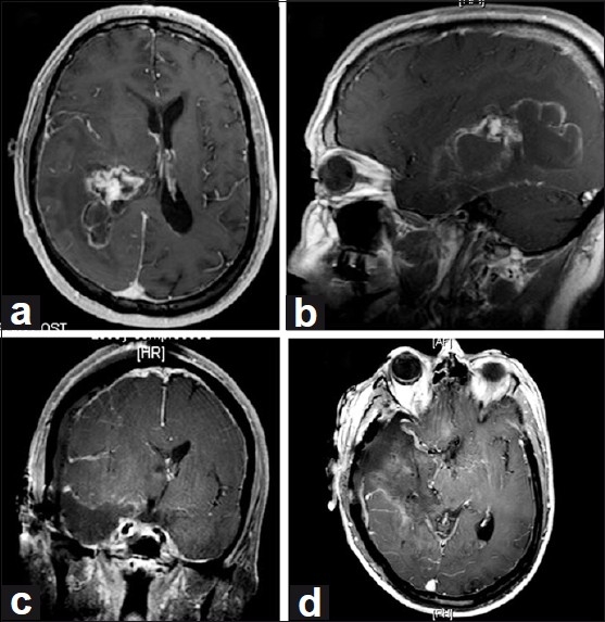
Background: The technique of sub-pial resection, first described in the early 1900s, was later refined by Penfield and Jasper for removal of supratentorial epileptic cortex. This technique has not been widely adopted for intrinsic tumor resection, for which the most widely used technique involves piecemeal aspiration of the tumor. This technique of "staying within the tumor" results in persistent bleeding, with obscuration of the tumor/brain interface, potentially yielding less than satisfactory results. In our experience, the sub-pial technique is useful for resections of supratentorial intrinsic tumor. We report the use of sub-pial resection technique and present illustrative cases.
Methods: The sub-pial resection technique is described along with important clinical decision-making guidelines. Representative cases are presented to discuss application of the sub-pial technique and to demonstrate surgical results.
Results: The sub-pial technique preserves the pia during cortical resections and makes it easier to protect and identify normal anatomy, including sulci, gyri, cranial nerves, and major vascular structures. This reduces bleeding, making surgery safer and more efficient. In most cases, an en bloc resection can be accomplished, permitting more accurate histopathology and more extensive tissue acquisition for research purposes.
Conclusion: The sub-pial technique can be incorporated into strategies for supratentorial intrinsic tumor resections, including temporal, frontal, occipital, and insular tumors, at para-Sylvian or para-insular-sulcus locations.
Keywords: En bloc resection; astrocytoma; neurosurgical procedures; sub-pial resection; supratentorial intrinsic tumor.
Figures


Similar articles
-
Comparative risk of leptomeningeal dissemination of cancer after surgery or stereotactic radiosurgery for a single supratentorial solid tumor metastasis.Neurosurgery. 2009 Apr;64(4):664-74; discussion 674-6. doi: 10.1227/01.NEU.0000341535.53720.3E. Neurosurgery. 2009. PMID: 19197219
-
Surgical resection of intrinsic insular tumors.Neurosurgery. 2005 Jul;57(1 Suppl):176-83; discussion 176-83. doi: 10.1227/01.neu.0000163603.70972.ab. Neurosurgery. 2005. PMID: 15987586 Review.
-
Neuronavigated Fiber Dissection with Pial Preservation: Laboratory Model to Simulate Opercular Approaches to Insular Tumors.World Neurosurg. 2017 Feb;98:239-242. doi: 10.1016/j.wneu.2016.10.020. Epub 2016 Oct 17. World Neurosurg. 2017. PMID: 27765721
-
Surgical resection of intrinsic insular tumors: complication avoidance.J Neurosurg. 2001 Oct;95(4):638-50. doi: 10.3171/jns.2001.95.4.0638. J Neurosurg. 2001. PMID: 11596959
-
Surgical and endovascular flow disconnection of intracranial pial single-channel arteriovenous fistulae.Neurosurgery. 2001 Dec;49(6):1351-63; discussion 1363-4. doi: 10.1097/00006123-200112000-00011. Neurosurgery. 2001. PMID: 11846934 Review.
Cited by
-
Perilesional Resection of Glioblastoma Is Independently Associated With Improved Outcomes.Neurosurgery. 2020 Jan 1;86(1):112-121. doi: 10.1093/neuros/nyz008. Neurosurgery. 2020. PMID: 30799490 Free PMC article.
-
Continuous monitoring of surgical bimanual expertise using deep neural networks in virtual reality simulation.NPJ Digit Med. 2022 Apr 26;5(1):54. doi: 10.1038/s41746-022-00596-8. NPJ Digit Med. 2022. PMID: 35473961 Free PMC article.
-
The role of Lobectomy in Glioblastoma management: A Retrospective series.Brain Spine. 2025 Jun 18;5:104305. doi: 10.1016/j.bas.2025.104305. eCollection 2025. Brain Spine. 2025. PMID: 40678087 Free PMC article.
-
Effect of Artificial Intelligence Tutoring vs Expert Instruction on Learning Simulated Surgical Skills Among Medical Students: A Randomized Clinical Trial.JAMA Netw Open. 2022 Feb 1;5(2):e2149008. doi: 10.1001/jamanetworkopen.2021.49008. JAMA Netw Open. 2022. PMID: 35191972 Free PMC article. Clinical Trial.
-
Safety and efficacy of concomitant chemotherapeutic wafers and iodine-125 seeds for recurrent glioblastoma.Surg Neurol Int. 2012;3:137. doi: 10.4103/2152-7806.103644. Epub 2012 Nov 20. Surg Neurol Int. 2012. PMID: 23230518 Free PMC article.
References
-
- Gil-Robles S, Duffau H. Surgical management of World Health Organization Grade II gliomas in eloquent areas: The necessity of preserving a margin around functional structures. Neurosurg Focus. 2010;28:E8. - PubMed
-
- Grant GA, Farrell D, Silbergeld DL. Continuous somatosensory evoked potential monitoring during brain tumor resection.Report of four cases and review of the literature. J Neurosurg. 2002;97:709–13. - PubMed
-
- Hall WA, Truwit CL. Intraoperative magnetic resonance imaging. Acta Neurochir Suppl. 2011;109:119–29. - PubMed
LinkOut - more resources
Full Text Sources
Research Materials
