. 2012 Feb 27:8:14.
doi: 10.1186/1744-8069-8-14.

The expression of spinal methyl-CpG-binding protein 2, DNA methyltransferases and histone deacetylases is modulated in persistent pain states

Affiliations

The expression of spinal methyl-CpG-binding protein 2, DNA methyltransferases and histone deacetylases is modulated in persistent pain states

Keri K Tochiki et al. Mol Pain. .

Abstract

Background: DNA CpG methylation is carried out by DNA methyltransferases and induces chromatin remodeling and gene silencing through a transcription repressor complex comprising the methyl-CpG-binding protein 2 (MeCP2) and a subset of histone deacetylases. Recently, we have found that MeCP2 activity had a crucial role in the pattern of gene expression seen in the superficial dorsal horn rapidly after injection of Complete Freund's Adjuvant (CFA) in the rat ankle joint. The aim of the present study was to analyse the changes in expression of MeCP2, DNA methyltransferases and a subset of histone deacetylases in the superficial dorsal horn during the maintenance phase of persistent pain states. In this process, the cell specific expression of MeCP2 was also investigated.

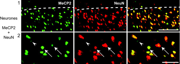
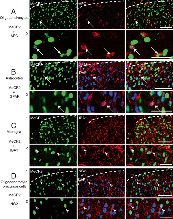
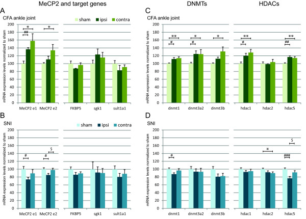
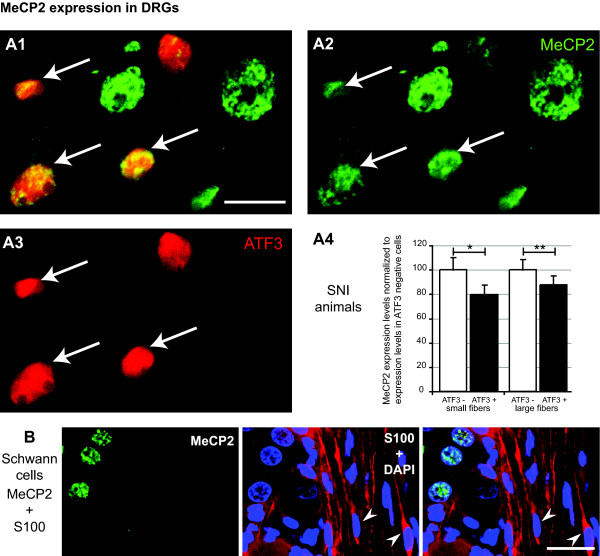
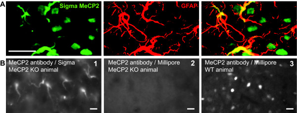
Results: Using immunohistochemistry, we found that neurones, oligodendrocytes and astrocytes expressed MeCP2. Microglia, oligodendrocyte precursor cells and Schwann cells never showed any positive stain for MeCP2. Quantitative analyses showed that MeCP2 expression was increased in the superficial dorsal horn 7 days following CFA injection in the ankle joint but decreased 7 days following spared nerve injury. Overall, the expression of DNA methyltransferases and a subset of histone deacetylases followed the same pattern of expression. However, there were no significant changes in the expression of the MeCP2 targets that we had previously shown are regulated in the early time points following CFA injection in the ankle joint. Finally, the expression of MeCP2 was also down regulated in damaged dorsal root ganglion neurones following spared nerve injury.

Conclusion: Our results strongly suggest that changes in chromatin compaction, regulated by the binding of MeCP2 complexes to methylated DNA, are involved in the modulation of gene expression in the superficial dorsal horn and dorsal root ganglia during the maintenance of persistent pain states.


Figures

Figure 1
Figure 1
All neurones express MeCP2 in the rat superficial dorsal horn. Confocal images of rat superficial dorsal horn sections. Colocalization of MeCP2 (green; Millipore antibody) and NeuN (red). MeCP2 can be seen within the nucleus of all neurones (arrows). However, some MeCP2 staining is clearly non-neuronal (arrow heads). Pictures show single focal plane. Scale bars, 1) 50 μm and 2) 20 μm.
Figure 2
Figure 2
MeCP2 is expressed in oligodendrocytes and astrocytes, but not in microglia or oligodendrocyte precursor cells, in the rat superficial dorsal horn. Confocal images of rat superficial dorsal horn sections. A, Colocalization of MeCP2 (green; Millipore antibody) and APC (red). MeCP2 can be seen within the nucleus of oligodendrocytes. B, Colocalization of MeCP2 (green; Millipore antibody), GFAP (red) and DAPI, a nuclear marker (blue). MeCP2 can be seen within the nucleus of astrocytes. C, Expression of MeCP2 (green; Millipore antibody) and IBA1 (red). MeCP2 stain cannot be seen within microglia. D, Expression of MeCP2 (green; Millipore antibody), NG2 (red) and DAPI, a nuclear marker (blue). MeCP2 stain cannot be seen within OPCs. Pictures show single focal plane. Scale bars, 1) 50 μm and 2) 20 μm.
Figure 3
Figure 3
Expression of MeCP2, DNMTs and HDACs increases within the dorsal horn 7 days following CFA injection in the ankle joint but decreases 7 days following SNI surgery. The expression of MeCP2, downstream targets, DNMTs and HDACs was quantified by RT-qPCR. Y axis represents mRNA expression as a percentage of control (sham animals). Data show mean ± SEM. *: P < 0.05 and ** P < 0.01 sham vs contra; # P < 0.05, ## P < 0.01 and ###: P < 0.001 sham vs ipsi; $ P < 0.05 ipsi vs contra. CFA: N = 5 in each group and SNI: N = 7 in each group.
Figure 4
Figure 4
MeCP2 expression is reduced within damaged DRG neurons following SNI surgery. A1 to A3, Images of DRG sections 7 days following SNI surgery. Colocalization of MeCP2 (green; Millipore antibody) and ATF3 (red) in DRG neurones. Arrow indicates neurones expressing both MeCP2 and ATF3. Scale bar, 20 μm. A4, Measure of MeCP2 expression in ATF3 positive neurons using confocal microscopy. MeCP2 expression was quantified by measuring staining intensity and normalized to MeCP2 expression in ATF3 negative cells within each sub-group (small and large fibers; cell body diameter < 25 μm and > 25 μm). B, Confocal images of rat dorsal root ganglion sections. Expression of MeCP2 (green; Millipore antibody), S100 (red) and DAPI, a nuclear marker (blue). Pictures show single focal plane. Scale bars, 50 μm.
Figure 5
Figure 5
MeCP2 antibody from Sigma wrongly labels astrocytic processes in the superficial dorsal horn. A, Colocalization of MeCP2 (green; Sigma antibody) and astrocytes (red) in the superficial dorsal horn of naïve rat. B, Images of dorsal horn sections of MeCP2 knock out animals (gift from A.Bird) stained with anti-MeCP2 antibody from Sigma (1) and from Millipore (2). (3) Dorsal horn of wild type animals (C57BL/6) stained with the Millipore anti-MeCP2 antibody. Scale bar, 20 μm.

References

    1. Valder CR, Liu JJ, Song YH, Luo ZD. Coupling gene chip analyses and rat genetic variances in identifying potential target genes that may contribute to neuropathic allodynia development. J Neurochem. 2003;87:560–573. doi: 10.1046/j.1471-4159.2003.02016.x. - DOI - PubMed
    1. Vega-Avelaira D, Geranton SM, Fitzgerald M. Differential regulation of immune responses and macrophage/neuron interactions in the dorsal root ganglion in young and adult rats following nerve injury. Mol Pain. 2009;5:70. doi: 10.1186/1744-8069-5-70. - DOI - PMC - PubMed
    1. Xiao HS, Huang QH, Zhang FX, Bao L, Lu YJ, Guo C. et al.Identification of gene expression profile of dorsal root ganglion in the rat peripheral axotomy model of neuropathic pain. Proc Natl Acad Sci USA. 2002;99:8360–8365. doi: 10.1073/pnas.122231899. - DOI - PMC - PubMed
    1. Yang HY, Mitchell K, Keller JM, Iadarola MJ. Peripheral inflammation increases Scya2 expression in sensory ganglia and cytokine and endothelial related gene expression in inflamed tissue. J Neurochem. 2007;103:1628–1643. doi: 10.1111/j.1471-4159.2007.04874.x. - DOI - PubMed
    1. Geranton SM, Morenilla-Palao C, Hunt SP. A role for transcriptional repressor methyl-CpG-binding protein 2 and plasticity-related gene serum- and glucocorticoid-inducible kinase 1 in the induction of inflammatory pain states. J Neurosci. 2007;27:6163–6173. doi: 10.1523/JNEUROSCI.1306-07.2007. - DOI - PMC - PubMed

Publication types

MeSH terms

LinkOut - more resources