. 2012 May 1;365(1):110-7.
doi: 10.1016/j.ydbio.2012.02.013. Epub 2012 Feb 18.

PDGF mediates TGFβ-induced migration during development of the spinous process

Affiliations

PDGF mediates TGFβ-induced migration during development of the spinous process

Ying Wang et al. Dev Biol. .

Abstract

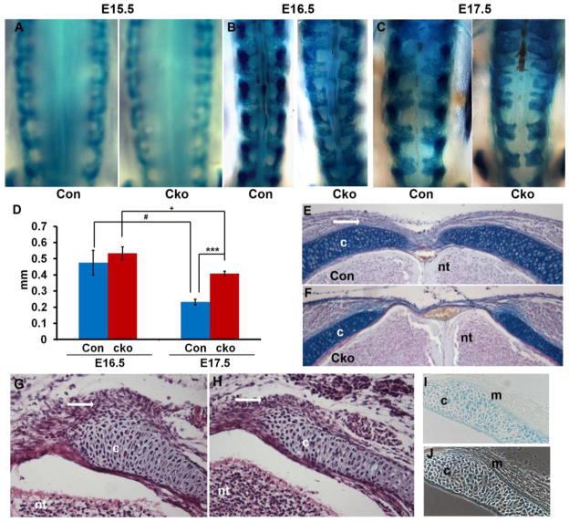
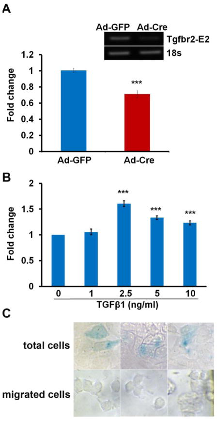
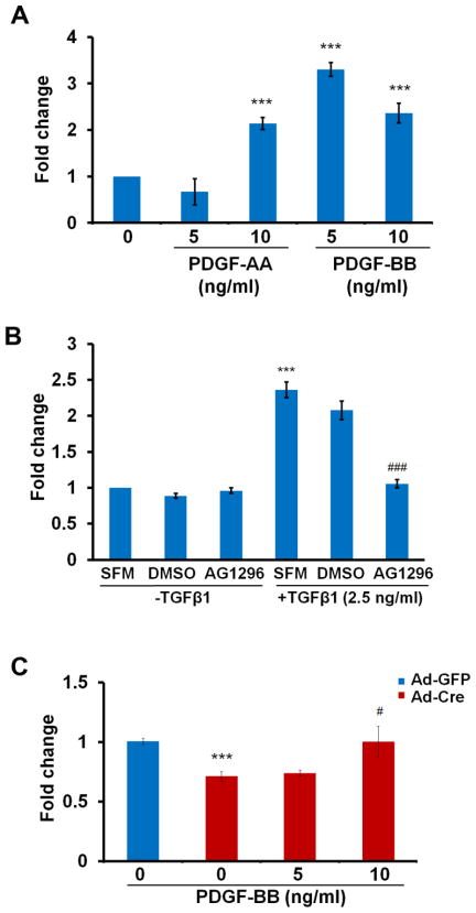
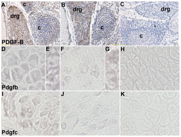
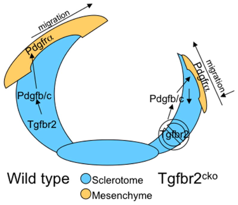
Mechanisms mediating closure of the dorsal vertebrae are not clear. Previously, we showed that deletion of TGFβ type II receptor (Tgfbr2) in sclerotome in mice results in failure in the formation of the spinous process, mimicking spina bifida occulta, a common malformation in humans. In this study, we aimed to determine whether missing dorsal structures in Tgfbr2 mutant mice were due to defects in mesenchymal migration and to clarify mechanism of TGFβ-mediated migration. First, we showed that gross alterations in dorsal vertebrae were apparent by E16.5days in Tgfbr2 mutants. In addition, histological staining showed that the mesenchyme adjacent to the developing cartilage was thin compared to controls likely due to reduced proliferation and migration of these cells. Next, we used a chemotaxis migration assay to show that TGFβ promotes migration in mixed cultures of embryonic sclerotome and associated mesenchyme. TGFβ stimulated expression of PDGF ligands and receptors in the cultures and intact PDGF signaling was required for TGFβ-mediated migration. Since PDGF ligands are expressed in the sclerotome-derived cartilage where Tgfbr2 is deleted and the receptors are predominantly expressed in the adjacent mesenchyme, we propose that TGFβ acts on the sclerotome to regulate expression of PDGF ligands, which then act on the associated mesenchyme in a paracrine fashion to mediate proliferation, migration and subsequent differentiation of the adjacent sclerotome.


Figures

Figure 1
Figure 1. Disrupted formation of the dorsal vertebrae in Tgfbr2cko embryos
(A–C) Representative images of whole mount X-gal staining on control (Con) and Tgfbr2cko (Cko) embryos at E15.5, E16.5 and E17.5 days. Magnification: 25×. (D) Measurements of the gap between neural arches at the second lumbar region from E16.5 and E17.5 embryos. Data was analyzed by 2-way ANOVA; N=5; ***, P<0.001 [E17.5 Cko vs E17.5 Con]; #, P<0.05 [E17.5 Con vs E16.5 Con]; +, P<0.05 [E17.5 Cko vs E16.5 Cko]. (E, F) Alcian blue staining of sections from lumbar vertebrae of E17.5 control and Tgfbr2cko embryos. Dense mesenchyme dorsal to the neural arch is seen in the control (white arrow). Magnification: 10×. (G, H) H & E staining of cryosections of the lumbar region from E15.5 control and Tgfbr2cko embryos. Dense mesenchyme adjacent to the sclerotome-derived cartilage is visible surrounding the dorsal edge of the neural arch in control mice (G, white arrow). This mesenchyme is reduced or absent in mutants (H, white arrow). Magnification: 20×. (I) Cryosection of X-gal stained E15.5 Col2aCre+;Rosa26/+ embryo. Cre activity is seen as blue X-gal stain in sclerotome derived cells. Staining is not observed in the adjacent sclerotome-associated mesenchyme. (J) Phase contrast image of I to show unstained adjacent mesenchyme. Magnification: 20×. c: cartilage; nt: neural tube; m: sclerotome associated mesenchyme.
Figure 2
Figure 2. TGFβ regulates migration in sclerotome associated mesenchyme in culture
(A) Sclerotome and associated mesenchyme was isolated from E11.5 Tgfbr2fl/fl embryos and infected with adenovirus Cre (Ad-Cre) to delete Tgfbr2 or with adenovirus GFP (Ad-GFP) as a control. Loss of Tgfbr2 resulted in reduced basal migration. ***, P<0.001. RT-PCR in the upper panel shows the deletion of the Tgfbr2 allele using Ad-Cre, 100 MOI. (B) The migration assay was performed on sclerotome and associated mesenchymal cells isolated from E11.5 wild type embryos and treated with or without TGFβ in the bottom well. TGFβ stimulated migration. ***, P<0.001. (C) Cells were isolated from Col2aCre+;Rosa26/+ mice and used in the migration assay with TGFβ in the bottom well. Sclerotome derived cells were stained blue with X-gal, unstained cells represent sclerotome associated cells. The total nonmigrated cell population demonstrated both stained and unstained cells. Migrated cells at the bottom of the chamber were not stained. Images were selected from several wells for both total and migrated cells. Magnification: 40×.
Figure 3
Figure 3. PDGF ligands plays an important role in TGFβ mediated cell migration
(A) Cells were isolated from wild type embryos and were either treated or not treated with PDGF-AA or PDGF-BB at the bottom of the well. Both PDGF ligands stimulated cell migration. ***, P<0.001. (B) Cells from wild type embryos were pre-treated with AG1296 (25 μM) for 30 min. before being placed in the migration assay with or without TGFβ in the bottom well. DMSO is the solvent control. SFM is the serum free medium control. TGFβ-stimulated migration was blocked by the PDGFR antagonist. ***, P<0.001 [SFM +TGFβ1 vs SFM-TGFβ1]; ###, P<0.001 [AG1296+TGFβ1 vs DMSO +TGFβ1]. (C) Cells were isolated from Tgfbr2fl/fl embryos and infected with Ad-Cre to delete Tgfbr2 or with Ad-GFP as a control. The cells were placed in the migration assay with or without PDGF-BB in the bottom well. PDGF-BB stimulated migration in Tgfbr2 deleted cells. ***, P<0.001 [Ad-Cre vs Ad-GFP]; #, P<0.01[Ad-Cre+PDGF-BB 10ng/ml vs Ad-Cre]. Representative of two separate experiments is shown.
Figure 4
Figure 4. PDGF-B and PDGF-C expression in Tgfbr2cko mice
(A–C) Immunostaining of PDGF-B protein on sections from control (A) and Tgfbr2cko (B) embryos at E15.5 days. Positive staining is seen as a brown DAB substrate. Negative control without primary antibody is shown in (C). c: cartilage, drg: dorsal root ganglion, Magnification: 40×. (D–H) In situ hybridization to Pdgfb mRNA in control (D, E) and Tgfbr2cko (F, G) cartilage (D, F) and dorsal root ganglion (E, G). (I-K) In situ hybridization to Pdgfc mRNA in control (I) and Tgfbr2cko (J) cartilage. Digoxigenin labelled RNA hybridization is seen as a purple NBT/BCIP substrate. Hybridization to a labelled sense probe is used as the negative control (H, K).
Figure 5
Figure 5. Proposed model for the role of Tgfbr2 in development of the spinous process
We propose the following model for TGFβ action in the development of the spinous process. In wild type embryos (left), TGFβ acts on its receptor in cartilage derived from sclerotome to stimulate PDGF ligand expression. PDGF ligands are then secreted and bind to PDGFRα on the adjacent mesenchyme. This paracrine activation of PDGF signaling promotes proliferation and migration of these cells eventually leading to the closure of the neural arches and formation of the spinous process. In Tgfbr2cko embryos (right), PDGF ligands are not made in sufficient amount to stimulate proliferation and migration of the adjacent mesenchyme. Alterations in the adjacent mesenchyme lead to failure in the formation of the spinous process.

Similar articles

Cited by

References

    1. Aase K, Abramsson A, Karlsson L, Betsholtz C, Eriksson U. Expression analysis of PDGF-C in adult and developing mouse tissues. Mechanisms of development. 2002;110:187–191. - PubMed
    1. Baffi MO, Moran MA, Serra R. Tgfbr2 regulates the maintenance of boundaries in the axial skeleton. Dev Biol. 2006;296:363–374. - PMC - PubMed
    1. Baffi MO, Slattery E, Sohn P, Moses HL, Chytil A, Serra R. Conditional deletion of the TGF-beta type II receptor in Col2a expressing cells results in defects in the axial skeleton without alterations in chondrocyte differentiation or embryonic development of long bones. Dev Biol. 2004;276:124–142. - PubMed
    1. Barrientos S, Stojadinovic O, Golinko MS, Brem H, Tomic-Canic M. Growth factors and cytokines in wound healing. Wound Repair Regen. 2008;16:585–601. - PubMed
    1. Bornfeldt KE, Raines EW, Graves LM, Skinner MP, Krebs EG, Ross R. Platelet-derived growth factor. Distinct signal transduction pathways associated with migration versus proliferation. Ann N Y Acad Sci. 1995;766:416–430. - PubMed

Publication types

MeSH terms

Substances

LinkOut - more resources