Atoh1, an essential transcription factor in neurogenesis and intestinal and inner ear development: function, regulation, and context dependency
- PMID: 22370966
- PMCID: PMC3346892
- DOI: 10.1007/s10162-012-0317-4
Atoh1, an essential transcription factor in neurogenesis and intestinal and inner ear development: function, regulation, and context dependency
Abstract
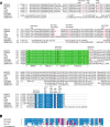
Atoh1 (also known as Math1, Hath1, and Cath1 in mouse, human, and chicken, respectively) is a proneural basic helix-loop-helix (bHLH) transcription factor that is required in a variety of developmental contexts. Atoh1 is involved in differentiation of neurons, secretory cells in the gut, and mechanoreceptors including auditory hair cells. Together with the two closely related bHLH genes, Neurog1 and NeuroD1, Atoh1 regulates neurosensory development in the ear as well as neurogenesis in the cerebellum. Atoh1 activity in the cochlea is both necessary and sufficient to drive auditory hair cell differentiation, in keeping with its known role as a regulator of various genes that are markers of terminal differentiation. Atoh1 is known in other fields as an oncogene and a tumor suppressor involved in regulation of cell cycle control and apoptosis. Aberrant Atoh1 activity in adult tissue is implicated in cancer progression, specifically in medullablastoma and adenomatous polyposis carcinoma. We demonstrate through protein sequence comparison that Atoh1 contains conserved phosphorylation sites outside the bHLH domain, which may allow regulation through post-translational modification. With such diverse roles, tight regulation of Atoh1 at both the transcriptional and protein level is essential.
Figures


Similar articles
-
The Role of Atonal Factors in Mechanosensory Cell Specification and Function.Mol Neurobiol. 2015 Dec;52(3):1315-1329. doi: 10.1007/s12035-014-8925-0. Epub 2014 Oct 23. Mol Neurobiol. 2015. PMID: 25339580 Free PMC article.
-
Neurod1 suppresses hair cell differentiation in ear ganglia and regulates hair cell subtype development in the cochlea.PLoS One. 2010 Jul 22;5(7):e11661. doi: 10.1371/journal.pone.0011661. PLoS One. 2010. PMID: 20661473 Free PMC article.
-
Atoh1: landscape for inner ear cell regeneration.Curr Gene Ther. 2014;14(2):101-11. doi: 10.2174/1566523214666140310143407. Curr Gene Ther. 2014. PMID: 24611726 Review.
-
Expression of Neurog1 instead of Atoh1 can partially rescue organ of Corti cell survival.PLoS One. 2012;7(1):e30853. doi: 10.1371/journal.pone.0030853. Epub 2012 Jan 24. PLoS One. 2012. PMID: 22292060 Free PMC article.
-
Development in the Mammalian Auditory System Depends on Transcription Factors.Int J Mol Sci. 2021 Apr 18;22(8):4189. doi: 10.3390/ijms22084189. Int J Mol Sci. 2021. PMID: 33919542 Free PMC article. Review.
Cited by
-
Characterization of the transcriptome of nascent hair cells and identification of direct targets of the Atoh1 transcription factor.J Neurosci. 2015 Apr 8;35(14):5870-83. doi: 10.1523/JNEUROSCI.5083-14.2015. J Neurosci. 2015. PMID: 25855195 Free PMC article.
-
A single-cell atlas of chromatin accessibility in mouse organogenesis.Nat Cell Biol. 2024 Jul;26(7):1200-1211. doi: 10.1038/s41556-024-01435-6. Epub 2024 Jul 8. Nat Cell Biol. 2024. PMID: 38977846
-
Oncomodulin regulates spontaneous calcium signalling and maturation of afferent innervation in cochlear outer hair cells.J Physiol. 2023 Oct;601(19):4291-4308. doi: 10.1113/JP284690. Epub 2023 Aug 29. J Physiol. 2023. PMID: 37642186 Free PMC article.
-
Bicistronic gene transfer tools for delivery of miRNAs and protein coding sequences.Int J Mol Sci. 2013 Sep 5;14(9):18239-55. doi: 10.3390/ijms140918239. Int J Mol Sci. 2013. PMID: 24013374 Free PMC article.
-
Initiation of Supporting Cell Activation for Hair Cell Regeneration in the Avian Auditory Epithelium: An Explant Culture Model.Front Cell Neurosci. 2020 Nov 12;14:583994. doi: 10.3389/fncel.2020.583994. eCollection 2020. Front Cell Neurosci. 2020. PMID: 33281558 Free PMC article.
References
-
- Aguado-Llera D, Goormaghtigh E, Geest N, Quan XJ, Prieto A, Hassan BA, Gomez J, Neira JL. The basic helix-loop-helix region of human neurogenin 1 is a monomeric natively unfolded protein which forms a “fuzzy” complex upon DNA binding. Biochemistry. 2010;49:1577–1589. doi: 10.1021/bi901616z. - DOI - PubMed
-
- Akazawa C, Ishibashi M, Shimizu C, Nakanishi S, Kageyama R. A mammalian helix-loop-helix factor structurally related to the product of Drosophila proneural gene atonal is a positive transcriptional regulator expressed in the developing nervous system. J Biol Chem. 1995;270:8730–8738. doi: 10.1074/jbc.270.3.1342. - DOI - PubMed
-
- Alder J, Lee KJ, Jessell TM, Hatten ME (1999) Generation of cerebellar granule neurons in vivo by transplantation of BMP-treated neural progenitor cells. Nat Neurosci 2:535–540 - PubMed
Publication types
MeSH terms
Substances
Grants and funding
LinkOut - more resources
Full Text Sources
Other Literature Sources
Molecular Biology Databases

