. 2012 Aug;97(8):1131-5.
doi: 10.3324/haematol.2011.052456. Epub 2012 Feb 27.

Estradiol increases hematopoietic stem and progenitor cells independent of its actions on bone

Affiliations

Estradiol increases hematopoietic stem and progenitor cells independent of its actions on bone

Anett Illing et al. Haematologica. 2012 Aug.

Abstract

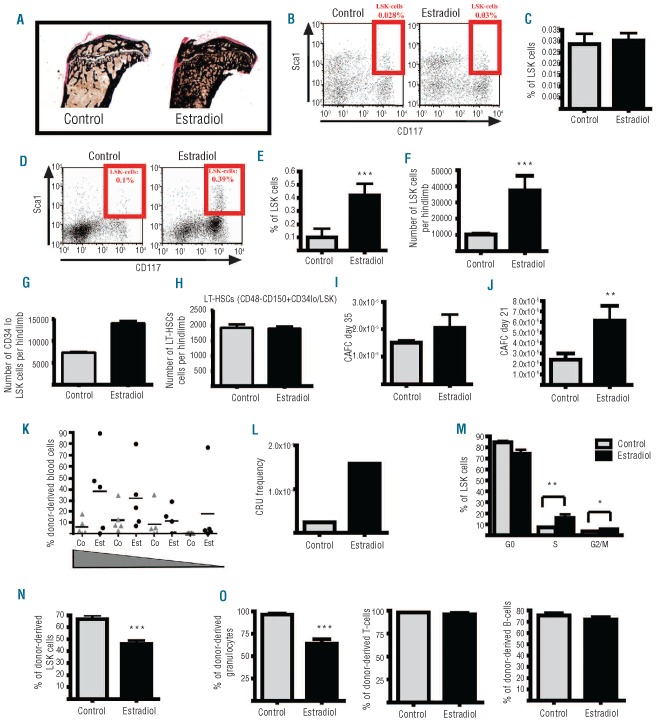
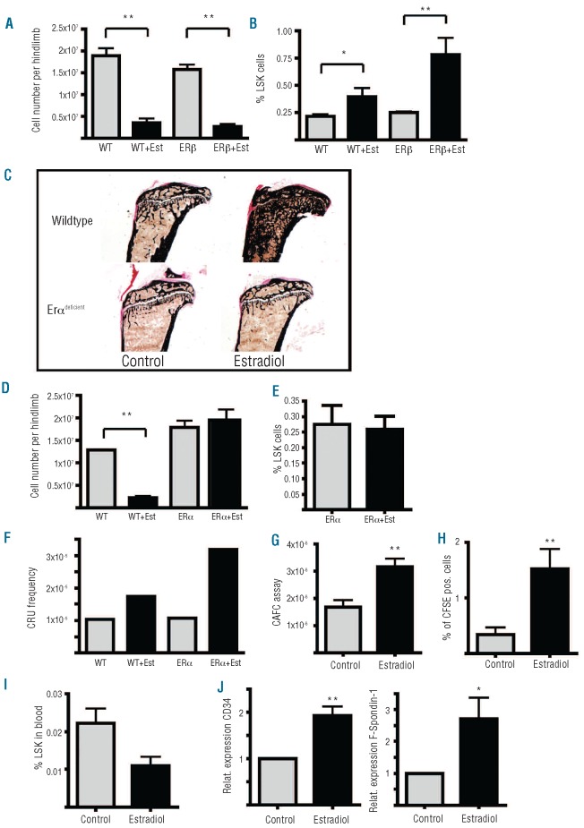
Hematopoietic stem and progenitor cells reside in vascular and endosteal niches in the bone marrow. Factors affecting bone remodeling were reported to influence numbers and mobilization of hematopoietic stem cells. We therefore analyzed the effects of estradiol acting anabolic on bone integrity. Here we observe that estradiol increases progenitor cell numbers in the vascular but not in the endosteal compartment independent of its estrogen receptor α-dependent anabolic bone effects. Hematopoietic progenitors capable of reconstituting lethally irradiated mice are increased by enhanced cell cycle entry, leading to a diminished long-term reconstitution potential after serial transplantation. We demonstrate that estradiol action on stromal cells potently favors hematopoietic progenitor/stem cell frequency accompanied by enhanced expression of cell adhesion molecules. Finally, estradiol treatment enhances retention of hematopoietic stem cells in the vascular niche of the bone marrow. We describe for the first time the mechanism of estrogen action on hematopoietic stem and progenitor cells.


Figures

Figure 1.
Figure 1.
Estradiol increases the bone mass in wild-type mice and functional HSPCs in the vascular niche of the BM by an enhanced entry into the cell cycle resulting in early exhaustion of LT-HSCs. (A) von Kossa staining for the calcium-content of the bones. Tibias of estradiol treated mice show increased calcium-content due to increased bone mass. (B) Representative dot blots of the bone-adherent hematopoietic cells stained for lineage-negative/Sca1-positive/cKit-positive cells (LSK-cells) isolated from estradiol and control treated mice. (C) Summarized percentages of LSK-cells in the fraction of bone-adherent hematopoietic cells in control and estradiol treated mice (n=5). (D) Representative dot blots of the cells of the vascular HSC-niche stained for lineage-negative/Sca1-positive/cKit-positive cells (LSK-cells) isolated from estradiol and control treated mice. (E) Summarized percentages of LSK-cells in the vascular niche of control and estradiol treated mice (n=5). (F) Absolute numbers of LSK-cells per hindlimb in the vascular niche from control and estradiol treated mice (n=5). (G, H) Absolute numbers of CD34−/lo LSK cells (G) and CD48-CD150+CD34−/lo LSK cells (H) per hindlimb in the vascular niche from control and estradiol treated mice (n=5). (I) Frequency of cobblestone area forming (CA-forming) colonies in the vascular niche of control and estradiol treated mice after 35 days of coculture. (J) Frequency of cobblestone area forming (CA-forming) colonies in the vascular niche of control and estradiol treated mice after 21 days of co-culture. (K) Percentages of donor-derived blood cells four months post transplantation with BM from control and estradiol treated mice in decreasing dilutions (dilutions: 540000, 180000, 60000, 20000 cells per mouse; n per dilution=5). (L) Calculated frequency of colony repopulating units (CRU), according to Poisson Statistics, 4 months post transplantation with BM from control and estradiol treated mice. (M) Distribution of LSK-cells throughout G0/G1, G2/M and the S-phase of the cell cycle after treatment of mice with estradiol (n=5). (N) Percentage of donor-derived (CD45.2-positive) LSK-cells after the third transplantation with BM from control and estradiol treated mice (n=10). (O) Percentage of donor-derived (CD45.2-positive) granulocytes (Gr1/CD11b-pos. cells) and donor-derived B (CD19-positive) and T cells (CD4/CD8-positive) after the third transplantation with BM from conrol and estradiol treated mice (n=10).
Figure 2.
Figure 2.
The ERα and the ERβ are redundant for the increase of vascular HSPCs by estradiol and estradiol increases the adhesion of HSPCs in the vascular niche by upregulation of adhesion molecules. (A) Absolute numbers of BM-cells per hindlimb in the vascular niche from control and estradiol treated wild-type and ERβ-ko mice (n=5). (B) Percentage of LSK-cells in the vascular niche of control and estradiol treated wild-type and ERβ-ko mice (n=5). (C) von Kossa staining for the calcium content of the bone on the tibia of control and estradiol treated wild-type and ERα-Runx2cre-mice. (D) Absolute numbers of BM-cells per hindlimb in the vascular niche from control and estradiol treated wild-type and ERα-ko mice (n=5). (E) Percentage of LSK-cells in the endosteal niche of control and estradiol treated ERα-ko mice (n=5). (F) Calculated frequency of colony repopulating units (CRU), using Poisson Statistics four months post transplantation with BM from control and estradiol treated wild-type and ERα-ko mice. CRU-calculation is based on the limiting-dilution-analysis (LDA), recipients received either 540000, 180000, 60000 or 20000 cells per mouse (n per dilution=5). (G) Frequency of cobblestone forming areas (CAs) formed by HSCs from the BM of wild-type mice after pre-treatment of FBMD1-feeder cells for 10 days with estradiol. (H) Percentage of CFSE-positive cells in the vascular niche of control and estradiol treated mice 15 h after injection of 1 million CFSE-labeled wild-type BM cells. (I) Relative mRNA-expression of F-Spondin 1 and CD34 in FBMD1-cells, ten days after estradiol treatment. (J) Percentage of LSK-cells in the blood of control and estradiol treated wild-type mice (n=5).

References

    1. Fuchs E, Tumbar T, Guasch G. Socializing with the neighbors: stem cells and their niche. Cell. 2004;116(6):769–78. - PubMed
    1. Sugiyama T, Kohara H, Noda M, Nagasawa T. Maintenance of the hematopoietic stem cell pool by CXCL12-CXCR4 chemokine signaling in bone marrow stromal cell niches. Immunity. 2006;25(6):977–88. - PubMed
    1. Calvi LM, Adams GB, Weibrecht KW, Weber JM, Olson DP, Knight MC, et al. Osteoblastic cells regulate the haematopoietic stem cell niche. Nature. 2003;425(6960):841–6. - PubMed
    1. Zhang J, Niu C, Ye L, Huang H, He X, Tong WG, et al. Identification of the haematopoietic stem cell niche and control of the niche size. Nature. 2003;425(6960):836–41. - PubMed
    1. Manolagas SC. From estrogen-centric to aging and oxidative stress: a revised perspective of the pathogenesis of osteoporosis. Endocr Rev. 2010;31(3):266–300. - PMC - PubMed

Publication types

MeSH terms