. 2013 Jul;34(7):1605-14.
doi: 10.1002/hbm.22013. Epub 2012 Feb 27.

Direct stimulation of the autonomic nervous system modulates activity of the brain at rest and when engaged in a cognitive task

Affiliations

Direct stimulation of the autonomic nervous system modulates activity of the brain at rest and when engaged in a cognitive task

Barbara Basile et al. Hum Brain Mapp. 2013 Jul.

Abstract

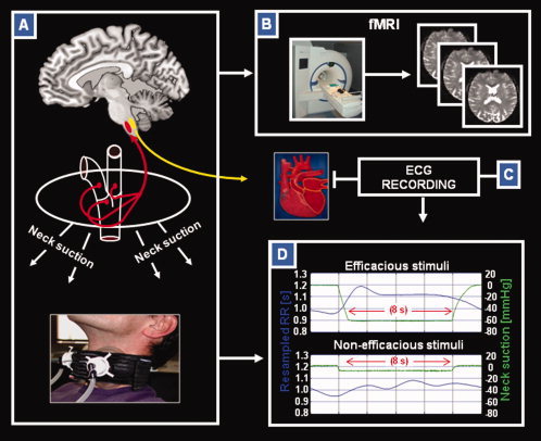
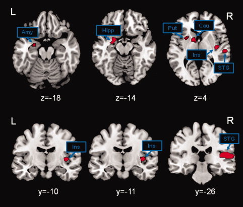
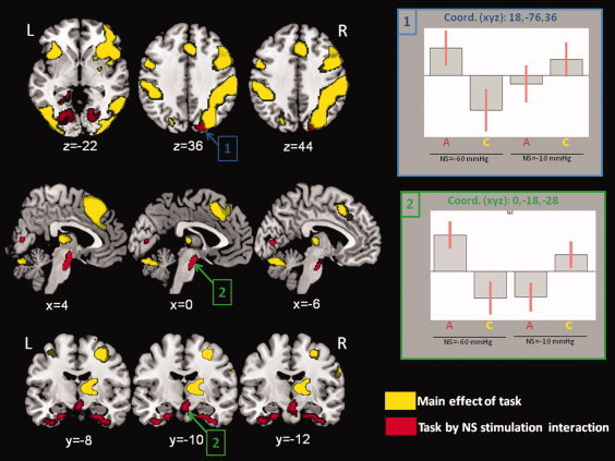
The effect of autonomic perturbation (AP) on the central nervous system functioning is still largely unknown. Using an automated neck suction device to stimulate the carotid mechanoreceptors in the carotid sinus (parasympathetic pathway), operated synchronously with functional magnetic resonance imaging (fMRI) acquisition, we investigated the effects of AP on the activity of the brain at rest and when engaged in a visuo-spatial attention task. ECG was always recorded to index changes in autonomic function. At rest, AP induced increased activation in the insula and in the amygdala, which have been previously associated with the autonomic control and emotion processing, as well as in the caudate nucleus and in the medial temporal cortex, both implicated in cognitive functions. Despite a preserved performance during visuo-spatial attention task, AP induced increased reaction times and a positive modulation on the activation of the right posterior parietal cortex, the occipital cortex, the periaquiductal gray, and nuclei of the brainstem. We speculate that this modulation of brain activity represents, at different anatomical levels, a compensation mechanism to maintain cognitive efficiency under parasympathetic stimulation, which is traditionally considered as the system for energy regain and storage. In conclusion, this study provides the first evidence of a dynamic interaction between AP and higher level functions in humans.


Figures

Figure 1
Figure 1
Neck suction (NS) technique used for fMRI investigation. Schematic illustration showing methods and devices used in the study. (A) Representation of the NS technique based on an MR compatible system (bottom); NS induces stimulation of the mechanoreceptors located in the carotid sinus bilaterally, thus producing an autonomic perturbation trough parasympathetic afferents (as detectable by cardiac effects); (B) 3T magnetic resonance scanner equipped for functional MRI (fMRI) experiments; (C) MR‐compliant system for ECG recording; (D) heart‐period stimulus response curve (shown in blue) and NS pressure (shown in green) for efficacious (−60 mm Hg) and nonefficacious (−10 mm Hg) stimuli. In this example, stimuli response curves were collected for one volunteer during experiment 1. Each curve is obtained as an average over 50 NS pulses at −60 mm Hg and 30 NS pulses at −10 mm Hg. Each stimulus has a duration of 8 s. The left axis refers to the heart period values, while the right axis reports the suction pressure applied to the neck. See text for further details.
Figure 2
Figure 2
fMRI paradigms. Schematic representation of the fMRI paradigms used to investigate the effect of autonomic perturbation on the brain in subjects at rest (experiment 1; panel A), and when engaged in performing a visuo‐spatial attention task (experiment 2; panel B). In experiment 1, subjects were required to lie still in the scanner, without thinking of anything in particular. Fifty efficacious (−60 mm Hg) and 30 nonefficacious (−10 mm Hg) neck suction (NS) pulses with a duration of 8 s each were randomly administered over 16 min of continuous fMRI data acquisition. Each NS pulse was followed by a random interpulse interval at atmospheric pressure ranging from 3.0 to 5.2 s. In experiment 2 (total fMRI acquisition = 16 min), the visuo‐spatial attention task included 32 blocks, with a duration of 30 s each, half of them requiring the active task (A = active), and half of them requiring a control condition (C = control). Each block started with instructions (duration = 3.2 s, followed by 0.8 s of black screen) and included the presentation of 14 stimuli (duration = 0.3 s each) separated from each other by intervals of black screen (duration = 1.7 s each). The effective intertrial interval was 2 s. The total number of trials was 896, with 224 repetitions for each of four trial‐types (active task under efficacious NS stimulation, active task under nonefficacious NS stimulation, control task under efficacious NS stimulation, and control task under nonefficacious NS stimulation). During the active condition, subjects were requested to press (as soon as possible) a button with the right index for items presenting with a symmetrical bisection of a colored horizontal line by a shorter vertical line and not to press in case of asymmetrical bisection. In the control condition, subjects were requested to press (as soon as possible) the same button when the items were presented in yellow and not to press in any other case. During task performance, NS was delivered in 60 s duration blocks, alternating half of them with a pressure of −60 mm Hg (efficacious stimulation), and the other half with a pressure of −10 mm Hg (nonefficacious stimulation). The active and control 30 s duration blocks were pseudo‐randomly administered in order to obtain half of them under efficacious and half under nonefficacious NS stimulation. Each block of NS consisted of 8 s duration pulses (either −60 or −10 mm Hg) interleaved with 4‐s duration intervals of suction release (cuffs at atmospheric pressure) to avoid the risk of accommodation for the mechanoreceptors. This allowed us to obtain a continuous stimulation of the mechanoreceptors over the entire block of efficacious NS stimulation. See text for further details.
Figure 3
Figure 3
Brain activation due to autonomic perturbation at rest. Brain activation for the comparison of efficacious greater than nonefficacious neck suction (NS) stimulation. Significant activations were observed in the amygdala, in the hippocampus, in the putamen, in the caudate nucleus, in the insula, and in the superior temporal gyrus. The inverse contrast (nonefficacious greater than efficacious NS stimulation) did not reveal any significant activation. Statistical threshold set to p‐FWE‐corr. <0.05 at cluster level. See text for further details. The xyz coordinates refer to MNI standard space. The xyz coordinates refer to MNI standard space. Abbreviations: amy, amygdala; hipp, hippocampus; put, putamen; cau, caudate nucleus; ins, insula; STG, superior temporal gyrus; R, right; L, left.
Figure 4
Figure 4
Changes in brain activation in the brain engaged in a visuo‐spatial attention task. Here, we show the main effect of task (yellow color) and the effect of task by neck suction (NS) stimulation interaction (red color). The main effect of task revealed the expected pattern of brain activation (right more than left side), including prefrontal, parietal and occipital cortex, the thalamus, and the cerebellum. The task by NS stimulation interaction revealed a positive modulation of activation in regions included in the main effect of task (posterior parietal cortex and occipital associative cortex) but also in other areas, such as the medial temporal pole bilaterally, the brainstem and the periaquiductal gray. Plots of signal changes across experimental conditions are shown for the cluster including the right posterior parietal cortex (plot 1) and for the cluster including the brainstem and periaquiductal gray (plot 2). Statistical threshold set to p‐FWE‐corr. < 0.05 at cluster level. The xyz coordinates refer to MNI standard space. See text for further details. Abbreviations: R, right; L, left; A = active task; C = control task.

References

    1. Allison T, Puce A, McCarthy G ( 2000): Social perception from visual cues: Role of the STS region. Trends Cogn Sci 4: 267–278. - PubMed
    1. Basile B, Mancini F, Macaluso E, Caltagirone C, Frackowiak RS, Bozzali M ( 2011): Deontological and altruistic guilt: Evidence for distinct neurobiological substrates. Hum Brain Mapp 32: 229–239. - PMC - PubMed
    1. Colivicchi F, Bassi A, Santini M, Caltagirone C ( 2004): Cardiac autonomic derangement and arrhythmias in right‐sided stroke with insular involvement. Stroke 35: 2094–2098. - PubMed
    1. Cooper VL, Hainsworth R ( 2009): Carotid baroreflex testing using the neck collar device. Clin Auton Res 19: 102–112. - PubMed
    1. Corbetta M, Akbudak E, Conturo TE, Snyder AZ, Ollinger JM, Drury HA, Linenweber MR, Petersen SE, Raichle ME, Van Essen DC, Shulman GL ( 1998): A common network of functional areas for attention and eye movements. Neuron 21: 761–773. - PubMed

Publication types

MeSH terms