

Selectivity filter gating in large-conductance Ca(2+)-activated K+ channels
- PMID: 22371364
- PMCID: PMC3289962
- DOI: 10.1085/jgp.201110748
Selectivity filter gating in large-conductance Ca(2+)-activated K+ channels
Abstract
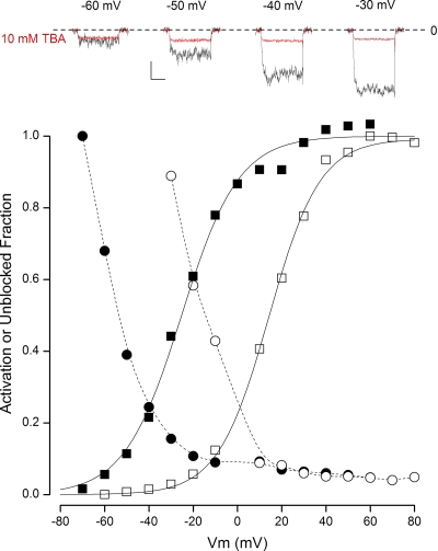
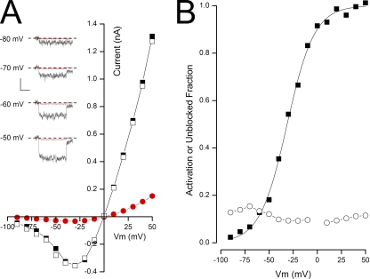
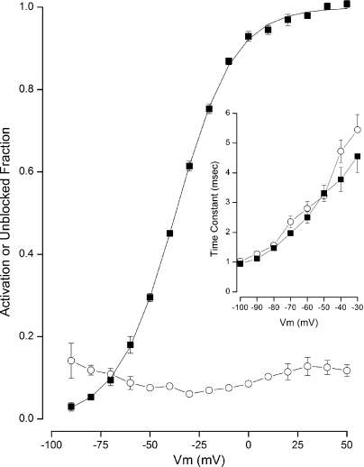
Membrane voltage controls the passage of ions through voltage-gated K (K(v)) channels, and many studies have demonstrated that this is accomplished by a physical gate located at the cytoplasmic end of the pore. Critical to this determination were the findings that quaternary ammonium ions and certain peptides have access to their internal pore-blocking sites only when the channel gates are open, and that large blocking ions interfere with channel closing. Although an intracellular location for the physical gate of K(v) channels is well established, it is not clear if such a cytoplasmic gate exists in all K(+) channels. Some studies on large-conductance, voltage- and Ca(2+)-activated K(+) (BK) channels suggest a cytoplasmic location for the gate, but other findings question this conclusion and, instead, support the concept that BK channels are gated by the pore selectivity filter. If the BK channel is gated by the selectivity filter, the interactions between the blocking ions and channel gating should be influenced by the permeant ion. Thus, we tested tetrabutyl ammonium (TBA) and the Shaker "ball" peptide (BP) on BK channels with either K(+) or Rb(+) as the permeant ion. When tested in K(+) solutions, both TBA and the BP acted as open-channel blockers of BK channels, and the BP interfered with channel closing. In contrast, when Rb(+) replaced K(+) as the permeant ion, TBA and the BP blocked both closed and open BK channels, and the BP no longer interfered with channel closing. We also tested the cytoplasmically gated Shaker K channels and found the opposite behavior: the interactions of TBA and the BP with these K(v) channels were independent of the permeant ion. Our results add significantly to the evidence against a cytoplasmic gate in BK channels and represent a positive test for selectivity filter gating.
© 2012 Thompson and Begenisich
Figures


Similar articles
-
State-dependent block of BK channels by synthesized shaker ball peptides.J Gen Physiol. 2006 Oct;128(4):423-41. doi: 10.1085/jgp.200609521. Epub 2006 Sep 11. J Gen Physiol. 2006. PMID: 16966472 Free PMC article.
-
Functional identification of ion binding sites at the internal end of the pore in Shaker K+ channels.J Physiol. 2003 May 15;549(Pt 1):107-20. doi: 10.1113/jphysiol.2002.038646. Epub 2003 Mar 28. J Physiol. 2003. PMID: 12665608 Free PMC article.
-
Unique inner pore properties of BK channels revealed by quaternary ammonium block.J Gen Physiol. 2004 Jul;124(1):43-57. doi: 10.1085/jgp.200409067. Epub 2004 Jun 14. J Gen Physiol. 2004. PMID: 15197222 Free PMC article.
-
Emerging issues of connexin channels: biophysics fills the gap.Q Rev Biophys. 2001 Aug;34(3):325-472. doi: 10.1017/s0033583501003705. Q Rev Biophys. 2001. PMID: 11838236 Review.
-
Molecular mechanisms of BK channel activation.Cell Mol Life Sci. 2009 Mar;66(5):852-75. doi: 10.1007/s00018-008-8609-x. Cell Mol Life Sci. 2009. PMID: 19099186 Free PMC article. Review.
Cited by
-
Cadmium-cysteine coordination in the BK inner pore region and its structural and functional implications.Proc Natl Acad Sci U S A. 2015 Apr 21;112(16):5237-42. doi: 10.1073/pnas.1500953112. Epub 2015 Apr 6. Proc Natl Acad Sci U S A. 2015. PMID: 25848005 Free PMC article.
-
Closed state-coupled C-type inactivation in BK channels.Proc Natl Acad Sci U S A. 2016 Jun 21;113(25):6991-6. doi: 10.1073/pnas.1607584113. Epub 2016 Jun 13. Proc Natl Acad Sci U S A. 2016. PMID: 27298368 Free PMC article.
-
Perspectives on: conformational coupling in ion channels: conformational coupling in BK potassium channels.J Gen Physiol. 2012 Dec;140(6):625-34. doi: 10.1085/jgp.201210849. J Gen Physiol. 2012. PMID: 23183698 Free PMC article.
-
Molecular mechanism of BK channel activation by the smooth muscle relaxant NS11021.J Gen Physiol. 2020 Jun 1;152(6):e201912506. doi: 10.1085/jgp.201912506. J Gen Physiol. 2020. PMID: 32221543 Free PMC article.
-
Selectivity filter ion binding affinity determines inactivation in a potassium channel.Proc Natl Acad Sci U S A. 2020 Nov 24;117(47):29968-29978. doi: 10.1073/pnas.2009624117. Epub 2020 Nov 5. Proc Natl Acad Sci U S A. 2020. PMID: 33154158 Free PMC article.
References
Publication types
MeSH terms
Substances
Grants and funding
LinkOut - more resources
Full Text Sources
Miscellaneous
