. 2012 Apr 1;188(7):3371-81.
doi: 10.4049/jimmunol.1100573. Epub 2012 Feb 27.

SHP-1 as a critical regulator of Mycoplasma pneumoniae-induced inflammation in human asthmatic airway epithelial cells

Affiliations

SHP-1 as a critical regulator of Mycoplasma pneumoniae-induced inflammation in human asthmatic airway epithelial cells

Ying Wang et al. J Immunol. .

Abstract

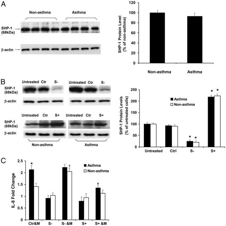
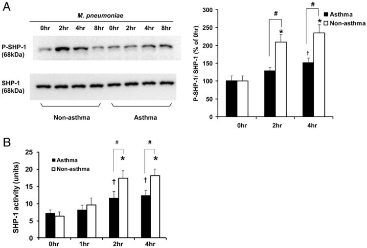
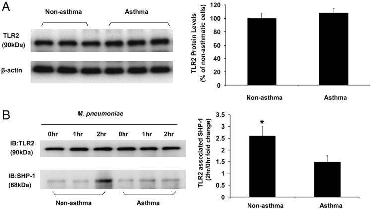
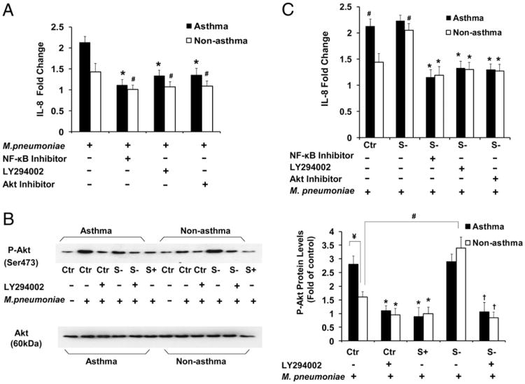
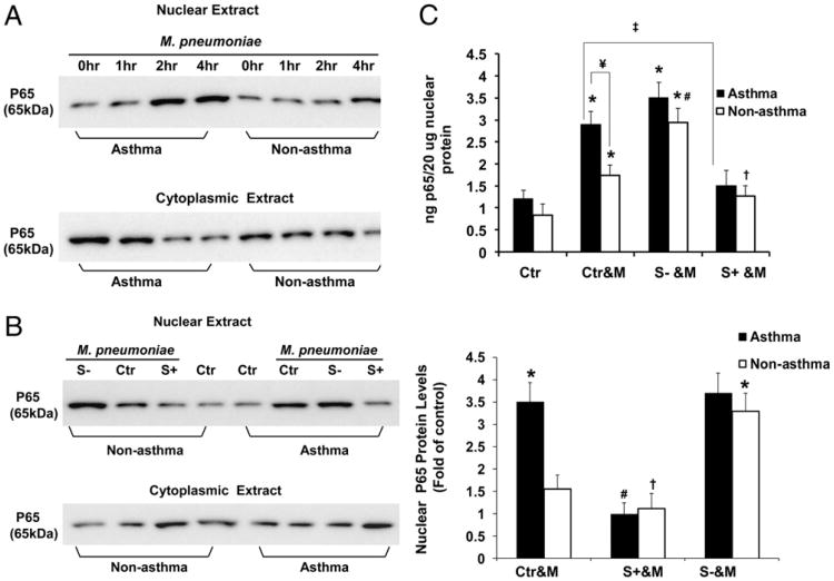
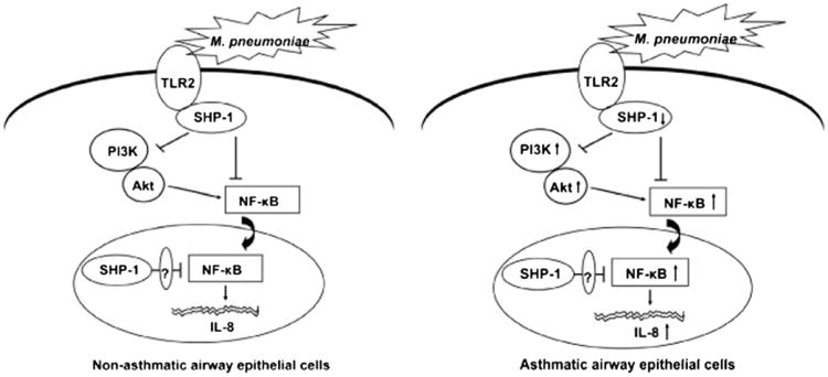
Asthma is a chronic inflammatory disease in which airway epithelial cells are the first line of defense against exposure of the airway to infectious agents. Src homology protein (SHP)-1, a protein tyrosine phosphatase, is a negative regulator of signaling pathways that are critical to the development of asthma and host defense. We hypothesize that SHP-1 function is defective in asthma, contributing to the increased inflammatory response induced by Mycoplasma pneumoniae, a pathogen known to exacerbate asthma. M. pneumoniae significantly activated SHP-1 in airway epithelial cells collected from nonasthmatic subjects by bronchoscopy with airway brushing but not in cells from asthmatic subjects. In asthmatic airway epithelial cells, M. pneumoniae induced significant PI3K/Akt phosphorylation, NF-κB activation, and IL-8 production compared with nonasthmatic cells, which were reversed by SHP-1 overexpression. Conversely, SHP-1 knockdown significantly increased IL-8 production and PI3K/Akt and NF-κB activation in the setting of M. pneumoniae infection in nonasthmatic cells, but it did not exacerbate these three parameters already activated in asthmatic cells. Thus, SHP-1 plays a critical role in abrogating M. pneumoniae-induced IL-8 production in nonasthmatic airway epithelial cells through inhibition of PI3K/Akt and NF-κB activity, but it is defective in asthma, resulting in an enhanced inflammatory response to infection.


Figures

Figure 1
Figure 1
Localization of endogenous SHP-1 protein in nonasthmatic and asthmatic human airway epithelial cells. Airway epithelial cells from nonasthmatic and asthmatic subjects were cultured at an air–liquid interface for 2 wk and immunostained using SHP-1–specific mAb. (A, D, G) SHP-1 (green) immunostaining. (B, E, H) Nuclear (red) immunostaining by 7-aminoactinomycin. (C, F, I) Merge. (G)–(I) are negative controls. SHP-1 was present in both cytoplasm and nucleus (A, D) but predominantly within the nucleus (C, F). Original magnification ×100.
Figure 2
Figure 2
IL-8 production in nonasthmatic and asthmatic airway epithelial cells infected with M. pneumoniae in the setting of SHP-1 knockdown and overexpression. (A) Immunoblot analysis of baseline SHP-1 expression in airway epithelial cells from nonasthmatic and asthmatic subjects. The data representing the percentage of densitometry values of nonasthmatic cells are expressed as mean ± SEM (right panel). (B) Immunoblot analysis of SHP-1 expression in airway epithelial cells without treatment (untreated), transduced with control rAAV only (Ctr), transduced with SHP-1–specific siRNA carrying rAAV to induce SHP-1 knockdown (S−), and infected with full-length SHP-1 cDNA carrying rAAV to induce SHP-1 overexpression (S+). The data representing the percentage of densitometry values of untreated cells are expressed as mean ± SEM (right panel). *p < 0.01 compared with untreated and control (Ctrl) in the same group. (C) Measurement of IL-8 production induced by M. pneumoniae in nonasthmatic and asthmatic airway epithelial cells with or without SHP-1 knockdown and overexpression. Data representing the fold change of IL-8 from control rAAV-transduced cells from the same group without mycoplasma infection are presented as mean ± SEM (n = 10 for nonasthma; n = 12 for asthma). Baseline SHP-1 expression was not different between asthmatic and nonasthmatic cells, and SHP-1 was knocked down and overexpressed similarly in nonasthmatic and asthmatic cells. IL-8 levels were higher in asthmatic cells infected with M. pneumoniae. SHP-1 overexpression reduced IL-8 production in asthmatic epithelial cells following mycoplasma infection. In the setting of SHP-1 knockdown, mycoplasma infection augmented IL-8 production in nonasthmatic, but not in asthmatic, cells.*,†p < 0.01 compared with Ctr&M of nonasthmatic cells; #p < 0.01 compared with Ctr&M or S− &M of asthmatic cells. Ctr&M, control rAAV-transduced airway epithelial cells treated with M. pneumoniae; S−, airway epithelial cells transduced with SHP-1–specific siRNA carrying rAAV to induce SHP-1 knockdown; S+, airway epithelial cells transduced with full-length SHP-1 cDNA carrying rAAV to induce SHP-1 overexpression; S− &M, airway epithelial cells with SHP-1 knocked down and infected by M. pneumoniae; S+ &M, airway epithelial cells with SHP-1 overexpressed and infected by M. pneumoniae.
Figure 3
Figure 3
M. pneumoniae infection in airway epithelial cells activates SHP-1. (A) M. pneumoniae infection of airway epithelial cells from non-asthmatic and asthmatic subjects resulted in time-course–dependent SHP-1 tyrosine phosphorylation. Airway epithelial cells without mycoplasma treatment (0 h) or infected with Mycoplasma for 2, 4, and 8 h were immunoprecipitated for SHP-1 and probed with anti-phosphotyrosine Ab 4G10 to detect P–SHP-1. Data representing P–SHP-1/SHP-1 of each time point compared with 0 h in nonasthmatic group are expressed as mean ± SEM, n = 5 (right panel). (B) Tyrosine phosphatase activity of immunoprecipitated SHP-1 from nonasthmatic and asthmatic airway epithelial cells without mycoplasma treatment (0 h) or infected with mycoplasma for 1, 2, or 4 h. *p < 0.01, p < 0.05, compared with 0 h in the same group; #p < 0.05 between nonasthmatic and asthmatic cells at the same time point. The data are presented as mean ± SEM (n = 10 for nonasthmatic subjects; n = 12 for asthmatic subjects). Although M. pneumoniae infection induced SHP-1 phosphorylation in asthmatic airway epithelial cells, it was significantly less than in nonasthmatic cells at 2 and 4 h following M. pneumoniae infection.
Figure 4
Figure 4
M. pneumoniae infection induced TLR2–SHP-1 dynamic association. (A) Immunoblot analysis of baseline TLR2 expression in airway epithelial cells from nonasthmatic and asthmatic subjects. The data representing the percentage of densitometry values of nonasthmatic cells are expressed as mean ± SEM (right panel). (B) TLR2-associated SHP-1 levels in airway epithelial cells was measured by coimmunoprecipitation. TLR2 was immunoprecipitated from airway epithelial cell lysates of nonasthmatic and asthmatic subjects without mycoplasma infection (0 h), infected with mycoplasma for 1 and 2 h. TLR2 immunoprecipitates were blotted with anti-TLR2 (top left panel) and anti–SHP-1 (bottom left panel) Ab. TLR2-associated SHP-1 at 2 h after M. pneumoniae infection is expressed as fold change of densitometry values from nonasthmatic cells without mycoplasma treatment (0 h) and was increased in nonasthmatic cells compared with asthmatic cells. The data are presented as mean ± SEM (n = 5). *p < 0.01 compared with asthmatic group.
Figure 5
Figure 5
SHP-1 regulates M. pneumoniae-induced IL-8 production through PI3K/Akt in airway epithelial cells from nonasthmatic and asthmatic subjects. (A) Fold change in IL-8 production in nonasthmatic and asthmatic airway epithelial cells with or without pretreatment with NF-κB inhibitor II (30 μM), LY294002 (25 μM), or Akt inhibitor (50 nM) for 30 min, followed by mycoplasma infection for 48 h. IL-8 levels are expressed as fold change from cells in the same group without mycoplasma infection. *p < 0.01, #p < 0.05, compared with mycoplasma-infected cells without inhibitor pretreatment in the same group. The data are presented as mean ± SEM (n = 10 for nonasthma; n = 12 for asthma). (B) Immunoblot analysis of Akt phosphorylation (P-Akt) in total cell lysates of nonasthmatic and asthmatic cells with or without mycoplasma infection for 2 h (left panel). The levels of P-Akt 2 h after M. pneumoniae infection in asthmatic and nonasthmatic cells, with or without SHP-1 knockdown or overexpression, are expressed as fold change of densitometry from cells in the same group transduced with control rAAV only (right panel). *p < 0.01, compared with control rAAV-transduced cells treated with mycoplasma in the same group; ¥,#p < 0.01, compared with control rAAV-transduced nonasthmatic cells treated with mycoplasma only; p < 0.01, compared with SHP-1 knockdown cells with mycoplasma infection only in the same group. Data are presented as mean ± SEM (n = 5). Mycoplasma-induced phosphorylation of Akt in asthmatic cells, which was significantly higher than that in nonasthmatic cells, was reversed by pretreatment with LY294002 and attenuated by SHP-1 overexpression. SHP-1 knockdown dramatically increased phosphorylation of Akt in nonasthmatic cells, but not in asthmatic cells, which was reversed by pretreatment with LY294002. (C) IL-8 fold change in SHP-1 knocked down nonasthmatic and asthmatic airway epithelial cells that were pretreated or not with NF-κB inhibitor II (30 μM), LY294002 (25 μM), or Akt inhibitor (50 nM) for 30 min and then infected with mycoplasma for 48 h. SHP-1 knockdown dramatically accentuated IL-8 production in nonasthmatic cells, but not in asthmatic cells, which were all significantly inhibited by these three inhibitors. *p < 0.01 compared with mycoplasma-infected “S−” cells in the same group, without pretreatment of inhibitors; #p < 0.01, compared with “Ctr” cells of nonasthma, without pretreatment of inhibitors. The data are presented as mean ± SEM (n = 10 for nonasthma; n = 12 for asthma). Ctr, control rAAV-transduced airway epithelial cells; S−, airway epithelial cells with SHP-1 knockdown; S+, airway epithelial cells with SHP-1 overexpression.
Figure 6
Figure 6
NF-κB activation by M. pneumoniae and inhibition by SHP-1 in airway epithelial cells from nonasthmatic and asthmatic subjects. (A) Immunoblot of cytoplasmic and nuclear protein lysates against p65 in nonasthmatic and asthmatic airway epithelial cells without mycoplasma infection (0 h) or challenged with mycoplasma for 1, 2, or 4 h. (B) Immunoblot of cytoplasmic and nuclear protein lysates against p65 in nonasthmatic and asthmatic airway epithelial cells with or without SHP-1 knockdown or overexpression and infected with mycoplasma for 2 h (left panel). The amount of nuclear p65 NF-κB at 2 h after M. pneumoniae infection is expressed as fold change of densitometry values from cells in the same group transduced with control rAAV only (right panel). *p < 0.01, compared with Ctr&M of nonasthmatic cells; #p < 0.01, p < 0.05, compared with Ctr&M in the same group. Data are presented as mean ± SEM (n = 5). (C) NF-κB activation was quantified in nonasthmatic and asthmatic airway epithelial cells, with or without SHP-1 knockdown or overexpression, 2 h after mycoplasma infection. The NF-κB DNA-binding activity is reported as nanograms of bound p65 protein/20 μg of nuclear extracts. *p < 0.01, compared with Ctr in the same group; #p < 0.01, S− &M versus Ctr&M of nonasthmatic cells; p < 0.05, S+ &M versus Ctr&M of nonasthmatic cells; p < 0.01, S+ &M versus Ctr&M of asthma; ¥p < 0.01, nonasthma versus asthma. Data are presented as mean ± SEM values (n = 10 for nonasthma; n = 12 for asthma). Mycoplasma induced NF-κB activation in asthmatic cells, which was significantly higher than that in nonasthmatic cells and was attenuated by SHP-1 overexpression. SHP-1 knockdown dramatically increased NF-κB activation in nonasthmatic cells but not in asthmatic cells. Ctr, control rAAV-transduced airway epithelial cells; Ctr&M, control rAAV-transduced cells with mycoplasma treatment; S−, airway epithelial cells with SHP-1 knockdown; S+, airway epithelial cells with SHP-1 overexpression; S− &M, airway epithelial cells with SHP-1 knocked down and infected by M. pneumoniae; S+ &M, airway epithelial cells with SHP-1 overexpressed and infected by M. pneumoniae.
Figure 7
Figure 7
Proposed model for the mechanism of SHP-1–mediated inhibition of M. pneumoniae-activated TLR2 signaling in normal and asthmatic airway epithelial cells. The ligation of TLR2 by M. pneumoniae initiates TLR2-mediated proinflammatory signaling pathway, resulting in the production of IL-8. M. pneumoniae binding to TLR2 activates and recruits SHP-1, which inhibits the nuclear translocation of NF-κB directly or through inhibition of PI3K/Akt abrogating NF-κB activation and subsequently prevents IL-8 production. In addition, the nuclear SHP-1 may also inhibit NF-κB function by certain nuclear mediators (left panel). In asthmatic airway epithelial cells, M. pneumoniae-induced SHP-1 activation is defective, which contributes to the increased activation of PI3K/Akt and NF-κB, as well as abundant IL-8 production (right panel).

Similar articles

Cited by

References

    1. Djukanović R, Wilson JW, Britten KM, Wilson SJ, Walls AF, Roche WR, Howarth PH, Holgate ST. Effect of an inhaled corticosteroid on airway inflammation and symptoms in asthma. Am Rev Respir Dis. 1992;145:669–674. - PubMed
    1. Wills-Karp M, Luyimbazi J, Xu X, Schofield B, Neben TY, Karp CL, Donaldson DD. Interleukin-13: central mediator of allergic asthma. Science. 1998;282:2258–2261. - PubMed
    1. Allegra L, Blasi F, Centanni S, Cosentini R, Denti F, Raccanelli R, Tarsia P, Valenti V. Acute exacerbations of asthma in adults: role of Chlamydia pneumoniae infection. Eur Respir J. 1994;7:2165–2168. - PubMed
    1. Berkovich S, Millian SJ, Snyder RD. The association of viral and mycoplasma infections with recurrence of wheezing in the asthmatic child. Ann Allergy. 1970;28:43–49. - PubMed
    1. Hahn DL, Dodge RW, Golubjatnikov R. Association of Chlamydia pneumoniae (strain TWAR) infection with wheezing, asthmatic bronchitis, and adult-onset asthma. JAMA. 1991;266:225–230. - PubMed

MeSH terms