Myenteric neurons of the mouse small intestine undergo significant electrophysiological and morphological changes during postnatal development
- PMID: 22371477
- PMCID: PMC3424759
- DOI: 10.1113/jphysiol.2011.225938
Myenteric neurons of the mouse small intestine undergo significant electrophysiological and morphological changes during postnatal development
Abstract
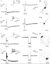
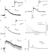
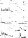
Organized motility patterns in the gut depend on circuitry within the enteric nervous system (ENS), but little is known about the development of electrophysiological properties and synapses within the ENS. We examined the electrophysiology and morphology of myenteric neurons in the mouse duodenum at three developmental stages: postnatal day (P)0, P10–11, and adult. Like adults, two main classes of neurons could be identified at P0 and P10–11 based on morphology: neurons with multiple long processes that projected circumferentially (Dogiel type II morphology) and neurons with a single long process. However, postnatal Dogiel type II neurons differed in several electrophysiological properties from adult Dogiel type II neurons. P0 and P10–11 Dogiel type II neurons exhibited very prominent Ca(2+)-mediated after depolarizing potentials (ADPs) following action potentials compared to adult neurons. Adult Dogiel type II neurons are characterized by the presence of a prolonged after hyperpolarizing potential (AHP), but AHPs were very rarely observed at P0. The projection lengths of the long processes of Dogiel type II neurons were mature by P10–11. Uniaxonal neurons in adults typically have fast excitatory postsynaptic potentials (fEPSPs, ‘S-type' electrophysiology) mainly mediated by nicotinic receptors. Nicotinic-fEPSPs were also recorded from neurons with a single long process at P0 and P10–11. However, these neurons underwent major developmental changes in morphology, from predominantly filamentous neurites at birth to lamellar dendrites in mature mice. Unlike Dogiel type II neurons, the projection lengths of neurons with a single long process matured after P10–11. Slow EPSPs were rarely observed in P0/P10–11 neurons. This work shows that, although functional synapses are present and two classes of neurons can be distinguished electrophysiologically and morphologically at P0, major changes in electrophysiological properties and morphology occur during the postnatal development of the ENS.
Figures


Comment in
-
The play is still being written on opening day: postnatal maturation of enteric neurons may provide an opening for early life mischief.J Physiol. 2012 May 15;590(10):2185-6. doi: 10.1113/jphysiol.2012.232769. J Physiol. 2012. PMID: 22589207 Free PMC article. No abstract available.
References
-
- Ambrogini P, Lattanzi D, Ciuffoli S, Agostini D, Bertini L, Stocchi V, Santi S, Cuppini R. Morpho-functional characterization of neuronal cells at different stages of maturation in granule cell layer of adult rat dentate gyrus. Brain Res. 2004;1017:21–31. - PubMed
-
- Baetge G, Gershon MD. Transient catecholaminergic (TC) cells in the vagus nerves and bowel of fetal mice – relationship to the development of enteric neurons. Dev Biol. 1989;132:189–211. - PubMed
-
- Bertrand PP, Kunze WAA, Furness JB, Bornstein JC. The terminals of myenteric intrinsic primary afferent neurons of the guinea-pig ileum are excited by 5-hydroxytryptamine acting at 5-hydroxytryptamine-3 receptors. Neuroscience. 2000;101:459–469. - PubMed
Publication types
MeSH terms
LinkOut - more resources
Full Text Sources
Other Literature Sources
Medical
Miscellaneous

