

Viruses and interactomes in translation
- PMID: 22371486
- PMCID: PMC3394946
- DOI: 10.1074/mcp.M111.014738
Viruses and interactomes in translation
Abstract
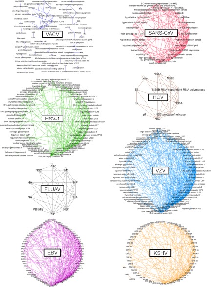
A decade of high-throughput screenings for intraviral and virus-host protein-protein interactions led to the accumulation of data and to the development of theories on laws governing interactome organization for many viruses. We present here a computational analysis of intraviral protein networks (EBV, FLUAV, HCV, HSV-1, KSHV, SARS-CoV, VACV, and VZV) and virus-host protein networks (DENV, EBV, FLUAV, HCV, and VACV) from up-to-date interaction data, using various mathematical approaches. If intraviral networks seem to behave similarly, they are clearly different from the human interactome. Viral proteins target highly central human proteins, which are precisely the Achilles' heel of the human interactome. The intrinsic structural disorder is a distinctive feature of viral hubs in virus-host interactomes. Overlaps between virus-host data sets identify a core of human proteins involved in the cellular response to viral infection and in the viral capacity to hijack the cell machinery for viral replication. Host proteins that are strongly targeted by a virus seem to be particularly attractive for other viruses. Such protein-protein interaction networks and their analysis represent a powerful resource from a therapeutic perspective.
Figures


Similar articles
-
Virus-host interactomes--antiviral drug discovery.Curr Opin Virol. 2012 Oct;2(5):614-21. doi: 10.1016/j.coviro.2012.09.003. Curr Opin Virol. 2012. PMID: 23057872 Free PMC article. Review.
-
Multimodal SARS-CoV-2 interactome sketches the virus-host spatial organization.Commun Biol. 2025 Mar 26;8(1):501. doi: 10.1038/s42003-025-07933-z. Commun Biol. 2025. PMID: 40140549 Free PMC article.
-
The domain landscape of virus-host interactomes.Biomed Res Int. 2014;2014:867235. doi: 10.1155/2014/867235. Epub 2014 Jun 4. Biomed Res Int. 2014. PMID: 24991570 Free PMC article.
-
A high-throughput yeast two-hybrid protocol to determine virus-host protein interactions.Methods Mol Biol. 2013;1064:1-15. doi: 10.1007/978-1-62703-601-6_1. Methods Mol Biol. 2013. PMID: 23996246 Free PMC article.
-
New horizons for antiviral drug discovery from virus-host protein interaction networks.Curr Opin Virol. 2012 Oct;2(5):606-13. doi: 10.1016/j.coviro.2012.09.001. Epub 2012 Sep 29. Curr Opin Virol. 2012. PMID: 23025912 Review.
Cited by
-
Local Action with Global Impact: Highly Similar Infection Patterns of Human Viruses and Bacteriophages.mSystems. 2016 Mar 8;1(2):e00030-15. doi: 10.1128/mSystems.00030-15. eCollection 2016 Mar-Apr. mSystems. 2016. PMID: 27822522 Free PMC article.
-
Beyond degree and betweenness centrality: Alternative topological measures to predict viral targets.PLoS One. 2018 May 24;13(5):e0197595. doi: 10.1371/journal.pone.0197595. eCollection 2018. PLoS One. 2018. PMID: 29795705 Free PMC article.
-
Mapping the SARS-CoV-2-Host Protein-Protein Interactome by Affinity Purification Mass Spectrometry and Proximity-Dependent Biotin Labeling: A Rational and Straightforward Route to Discover Host-Directed Anti-SARS-CoV-2 Therapeutics.Int J Mol Sci. 2021 Jan 7;22(2):532. doi: 10.3390/ijms22020532. Int J Mol Sci. 2021. PMID: 33430309 Free PMC article. Review.
-
Elucidating novel hepatitis C virus-host interactions using combined mass spectrometry and functional genomics approaches.Mol Cell Proteomics. 2014 Jan;13(1):184-203. doi: 10.1074/mcp.M113.030155. Epub 2013 Oct 29. Mol Cell Proteomics. 2014. PMID: 24169621 Free PMC article.
-
COVID-19 engages clinical markers for the management of cancer and cancer-relevant regulators of cell proliferation, death, migration, and immune response.Sci Rep. 2021 Mar 4;11(1):5228. doi: 10.1038/s41598-021-84780-y. Sci Rep. 2021. PMID: 33664395 Free PMC article.
References
-
- Flajolet M., Rotondo G., Daviet L., Bergametti F., Inchauspé G., Tiollais P., Transy C., Legrain P. (2000) A genomic approach of the hepatitis C virus generates a protein interaction map. Gene 242, 369–379 - PubMed
Publication types
MeSH terms
Substances
LinkOut - more resources
Full Text Sources
Miscellaneous
