. 2012 Apr 27;287(18):14873-9.
doi: 10.1074/jbc.M112.345751. Epub 2012 Feb 27.

Defective retinal depolarizing bipolar cells in regulators of G protein signaling (RGS) 7 and 11 double null mice

Affiliations

Defective retinal depolarizing bipolar cells in regulators of G protein signaling (RGS) 7 and 11 double null mice

Hoon Shim et al. J Biol Chem. .

Abstract

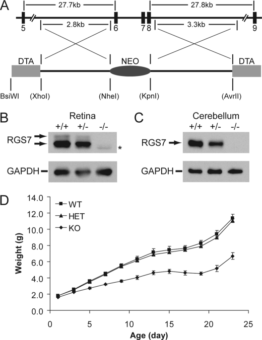
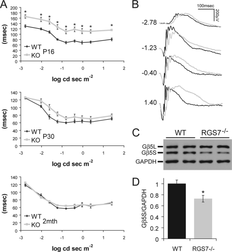
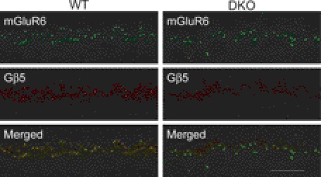
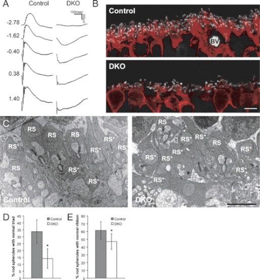
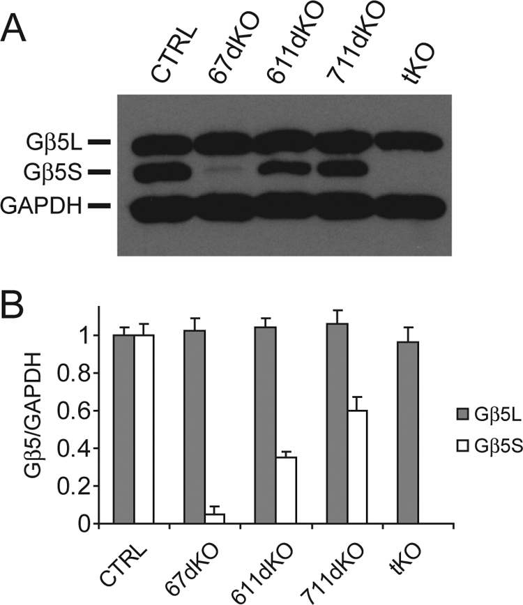
Two members of the R7 subfamily of regulators of G protein signaling, RGS7 and RGS11, are present at dendritic tips of retinal depolarizing bipolar cells (DBCs). Their involvement in the mGluR6/Gα(o)/TRPM1 pathway that mediates DBC light responses has been implicated. However, previous genetic studies employed an RGS7 mutant mouse that is hypomorphic, and hence the exact role of RGS7 in DBCs remains unclear. We have made a true RGS7-null mouse line with exons 6-8 deleted. The RGS7(-/-) mouse is viable and fertile but smaller in body size. Electroretinogram (ERG) b-wave implicit time in young RGS7(-/-) mice is prolonged at eye opening, but the phenotype disappears at 2 months of age. Expression levels of RGS6 and RGS11 are unchanged in RGS7(-/-) retina, but the Gβ5S level is significantly reduced. By characterizing a complete RGS7 and RGS11 double knock-out (711dKO) mouse line, we found that Gβ5S expression in the retinal outer plexiform layer is eliminated, as is the ERG b-wave. Ultrastructural defects akin to those of Gβ5(-/-) mice are evident in 711dKO mice. In retinas of mice lacking RGS6, RGS7, and RGS11, Gβ5S is undetectable, whereas levels of the photoreceptor-specific Gβ5L remain unchanged. Whereas RGS6 alone sustains a significant amount of Gβ5S expression in retina, the DBC-related defects in Gβ5(-/-) mice are caused solely by a combined loss of RGS7 and RGS11. Our data support the notion that the role of Gβ5 in the retina, and likely in the entire nervous system, is mediated exclusively by R7 RGS proteins.


Figures

FIGURE 1.
FIGURE 1.
RGS7−/− mice have a lower body weight. A, partial map of the mouse RGS7 gene, targeted exons, and the targeting construct containing negative and positive selection markers in DTA and NEO, respectively. Upon homologous recombination, exons 6–8 are replaced with the NEO marker, whereas the DTA markers are concurrently lost. Restriction enzyme sites in parentheses are present in the backbone pDND7-F vector and were introduced by PCR during the amplification of homology arms. B and C, Western blotting analysis of protein extracts derived from retina (20 μg, B) and cerebellum (10 μg, C) in littermate wild-type (+/+), heterozygous (+/−), and homozygous (−/−) RGS7 knock-out mice using anti-RGS6/7 (H-190, 1:2,000) antibody indicates the complete loss of RGS7 in RGS7−/− mice. Anti-GAPDH (1:50,000) serves as loading control. Asterisk denotes RGS6 signal picked up by the H-190 antibody. D, weights of RGS7+/+ (WT, n = 4), RGS7+/− (HET, n = 6), and RGS7−/− (KO, n = 4) mice recorded from P1 to P23 at 2-day intervals, showing that KO mice have an early noticeable smaller body weight (p < 0.01, P3–P23) compared with those of WT and HET littermates.
FIGURE 2.
FIGURE 2.
RGS7−/− mice exhibit an age-dependent delayed ERG b-wave implicit time as well as loss of Gβ5S protein in the retina. A, ERG b-wave implicit time, measured as the time difference between flash onset and the time b-wave is at its peak, was significantly delayed (*, p < 0.01) after eye opening at P16 in RGS7−/− mice (KO, n = 8) compared with WT littermate controls (n = 8). At P30, the KO (n = 6) implicit time delay was improved compared with WT (n = 6), showing a slight trend, but this delay was no longer statistically significant. At 2 months of age (2mth), the b-wave implicit time of KO (n = 8) mice is indistinguishable from that of WT animals (n = 6). Error bars are S.E. B, representative ERG traces from WT (black) and KO (gray) animals at P16 are shown with flash intensity indicated at left in unit of log cd s m−2. C and D, Gβ5S protein level in the retina was decreased in adult RGS7−/− mouse at P30. C, representative immunoblot shows the level of Gβ5S from two WT and two RGS7−/− mice retinal protein extracts (20 μg) using anti-Gβ5 (CT215, 1:2,000) and anti-GAPDH (1:50,000) antibodies. D, quantification of Gβ5S protein level, normalized to GAPDH signals, shows a significant 27 ± 7% reduction in the RGS7−/− (n = 6) versus WT (n = 4) mice (*, p < 0.01). Error bars are S.E.
FIGURE 3.
FIGURE 3.
Absence of Gβ5 immunoreactivity in mGluR6-containing puncta in retinas of 711dKO mice. Retinal sections from WT control and 711dKO (DKO) mice stained for metabotropic glutamate receptor 6 (mGluR6, 1:200) and Gβ5 (CT215, 1:250) demonstrate superimposed signals in the WT but absence of Gβ5 signal in the DKO. Scale bar, 10 μm.
FIGURE 4.
FIGURE 4.
RGS7 and RGS11 dKO mice have no ERG b-waves and contain ultrastructural defects in the retinal OPL. A, representative scotopic ERG responses from 2-month-old WT control and 711dKO (DKO) mice with flash intensity indicated at left in units of log cd s m−2. B, immunohistochemical staining of control and DKO retinal sections for CtBP2 (1:2,000, white) and PKCα (1:100,000, red) demonstrating reduction in rod bipolar cell dendritic arborization. BV, blood vessel. Scale bar, 5 μm. C, representative transmission electron microscopic images of the OPL from control and DKO mice. Scale bar, 2 μm. RS, rod spherule. Asterisk denotes abnormal structure. D and E, percentage of rod spherules with normal synaptic triads (D) and ribbons (E) in the DKO (n = 4, 80 images) versus control (n = 3, 65 images) retinas (*, p < 0.01). Error bars are S.D.
FIGURE 5.
FIGURE 5.
Gβ5S protein is undetectable in the RGS6, RGS7, and RGS11 tKO mouse retina. A, representative immunoblot showing the level of Gβ5L and Gβ5S from WT CTRL, 67dKO, 611dKO, 711dKO, and tKO mouse retinal extracts (15 μg) using anti-Gβ5 (CT215, 1:2,000) and anti-GAPDH (1:50,000) antibodies. B, quantification of Gβ5L and Gβ5S levels, normalized to GAPDH signal. Retinal Gβ5L protein levels do not differ across the various genotypes. However, 67dKO contains ∼5% of normal Gβ5S protein level (n = 3), 611dKO contains ∼35% of normal Gβ5S protein level (n = 3), 711dKO contains ∼60% of normal Gβ5S protein level (n = 3), and tKO has undetectable Gβ5S protein level (n = 3). Error bars are S.E.

Similar articles

Cited by

References

    1. Ross E. M., Wilkie T. M. (2000) GTPase-activating proteins for heterotrimeric G proteins: regulators of G protein signaling (RGS) and RGS-like proteins. Annu. Rev. Biochem. 69, 795–827 - PubMed
    1. Chen C. K., Eversole-Cire P., Zhang H., Mancino V., Chen Y. J., He W., Wensel T. G., Simon M. I. (2003) Instability of GGL domain-containing RGS proteins in mice lacking the G protein β-subunit Gβ5. Proc. Natl. Acad. Sci. U.S.A. 100, 6604–6609 - PMC - PubMed
    1. Cheever M. L., Snyder J. T., Gershburg S., Siderovski D. P., Harden T. K., Sondek J. (2008) Crystal structure of the multifunctional Gβ5-RGS9 complex. Nat. Struct. Mol. Biol. 15, 155–162 - PMC - PubMed
    1. Snow B. E., Krumins A. M., Brothers G. M., Lee S. F., Wall M. A., Chung S., Mangion J., Arya S., Gilman A. G., Siderovski D. P. (1998) A G protein γ subunit-like domain shared between RGS11 and other RGS proteins specifies binding to Gβ5 subunits. Proc. Natl. Acad. Sci. U.S.A. 95, 13307–13312 - PMC - PubMed
    1. Rao A., Dallman R., Henderson S., Chen C. K. (2007) Gβ5 is required for normal light responses and morphology of retinal ON-bipolar cells. J. Neurosci. 27, 14199–14204 - PMC - PubMed

Publication types