

Structure of factor H-binding protein B (FhbB) of the periopathogen, Treponema denticola: insights into progression of periodontal disease
- PMID: 22371503
- PMCID: PMC3339992
- DOI: 10.1074/jbc.M112.339721
Structure of factor H-binding protein B (FhbB) of the periopathogen, Treponema denticola: insights into progression of periodontal disease
Abstract
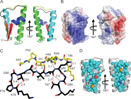
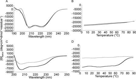
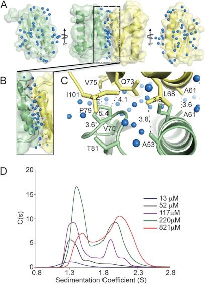
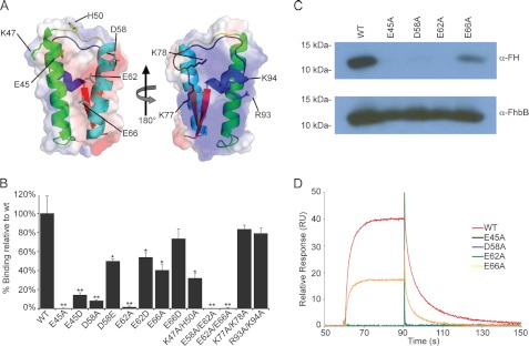
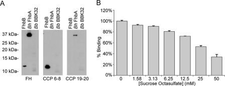
Periodontitis is the most common disease of microbial etiology in humans. Periopathogen survival is dependent upon evasion of complement-mediated destruction. Treponema denticola, an important contributor to periodontitis, evades killing by the alternative complement cascade by binding factor H (FH) to its surface. Bound FH is rapidly cleaved by the T. denticola protease, dentilisin. In this report, the structure of the T. denticola FH-binding protein, FhbB, was solved to 1.7 Å resolution. FhbB possesses a unique fold that imparts high thermostability. The kinetics of the FH/FhbB interaction were assessed using surface plasmon resonance. A K(D) value in the micromolar range (low affinity) was demonstrated, and rapid off kinetics were observed. Site-directed mutagenesis and sucrose octasulfate competition assays collectively indicate that the negatively charged face of FhbB binds within FH complement control protein module 7. This study provides significant new insight into the molecular basis of FH/FhbB interaction and advances our understanding of the role that T. denticola plays in the development and progression of periodontal disease.
Figures
Similar articles
-
Analysis of the complement sensitivity of oral treponemes and the potential influence of FH binding, FH cleavage and dentilisin activity on the pathogenesis of periodontal disease.Mol Oral Microbiol. 2014 Oct;29(5):194-207. doi: 10.1111/omi.12054. Epub 2014 Jun 3. Mol Oral Microbiol. 2014. PMID: 24815960 Free PMC article.
-
Analysis of a unique interaction between the complement regulatory protein factor H and the periodontal pathogen Treponema denticola.Infect Immun. 2009 Apr;77(4):1417-25. doi: 10.1128/IAI.01544-08. Epub 2009 Feb 9. Infect Immun. 2009. PMID: 19204088 Free PMC article.
-
Identification of the primary mechanism of complement evasion by the periodontal pathogen, Treponema denticola.Mol Oral Microbiol. 2011 Apr;26(2):140-9. doi: 10.1111/j.2041-1014.2010.00598.x. Epub 2010 Dec 3. Mol Oral Microbiol. 2011. PMID: 21375704 Free PMC article.
-
Treponema denticola major surface protein (Msp): a key player in periodontal pathogenicity and immune evasion.Arch Microbiol. 2025 Jan 18;207(2):36. doi: 10.1007/s00203-024-04223-w. Arch Microbiol. 2025. PMID: 39825920 Review.
-
Gene Regulation, Two Component Regulatory Systems, and Adaptive Responses in Treponema Denticola.Curr Top Microbiol Immunol. 2018;415:39-62. doi: 10.1007/82_2017_66. Curr Top Microbiol Immunol. 2018. PMID: 29026924 Review.
Cited by
-
Analysis of the antigenic determinants of the OspC protein of the Lyme disease spirochetes: Evidence that the C10 motif is not immunodominant or required to elicit bactericidal antibody responses.Vaccine. 2019 Apr 17;37(17):2401-2407. doi: 10.1016/j.vaccine.2019.02.007. Epub 2019 Mar 25. Vaccine. 2019. PMID: 30922701 Free PMC article.
-
Navigating infection by pathogenic spirochetes: The host-bacteria interface at the atomic level.Protein Sci. 2025 Jul;34(7):e70185. doi: 10.1002/pro.70185. Protein Sci. 2025. PMID: 40545683 Free PMC article. Review.
-
Development and optimization of OspC chimeritope vaccinogens for Lyme disease.Vaccine. 2020 Feb 18;38(8):1915-1924. doi: 10.1016/j.vaccine.2020.01.027. Epub 2020 Jan 17. Vaccine. 2020. PMID: 31959423 Free PMC article.
-
The Cross-Talk between Spirochetal Lipoproteins and Immunity.Front Immunol. 2014 Jun 30;5:310. doi: 10.3389/fimmu.2014.00310. eCollection 2014. Front Immunol. 2014. PMID: 25071771 Free PMC article. Review.
-
Hijacking Factor H for Complement Immune Evasion.Front Immunol. 2021 Feb 25;12:602277. doi: 10.3389/fimmu.2021.602277. eCollection 2021. Front Immunol. 2021. PMID: 33717083 Free PMC article. Review.
References
-
- Darveau R. P. (2010) Periodontitis. A polymicrobial disruption of host homeostasis. Nat. Rev. Microbiol. 8, 481–490 - PubMed
-
- Van Dyke T. E., Serhan C. N. (2003) Resolution of inflammation. A new paradigm for the pathogenesis of periodontal diseases. J. Dent. Res. 82, 82–90 - PubMed
-
- Paster B. J., Olsen I., Aas J. A., Dewhirst F. E. (2006) The breadth of bacterial diversity in the human periodontal pocket and other oral sites. Periodontol. 2000 42, 80–87 - PubMed
Publication types
MeSH terms
Substances
Associated data
- Actions
Grants and funding
LinkOut - more resources
Full Text Sources
Miscellaneous
