. 2012 Apr;158(4):1583-99.
doi: 10.1104/pp.112.194001. Epub 2012 Feb 27.

Subclassification and biochemical analysis of plant papain-like cysteine proteases displays subfamily-specific characteristics

Affiliations

Subclassification and biochemical analysis of plant papain-like cysteine proteases displays subfamily-specific characteristics

Kerstin H Richau et al. Plant Physiol. 2012 Apr.

Abstract

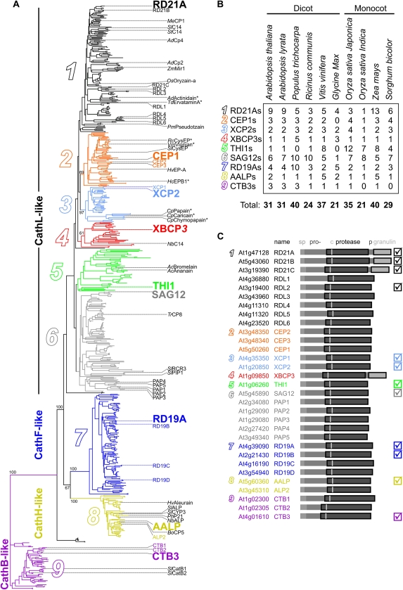
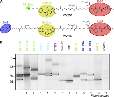
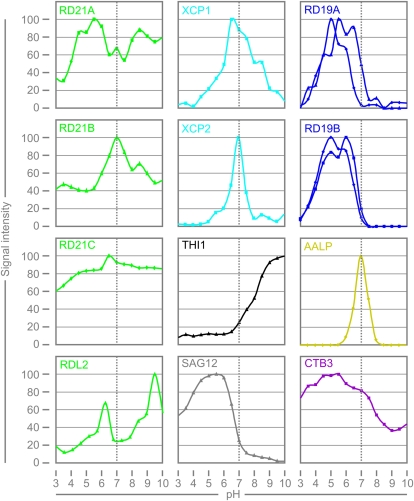
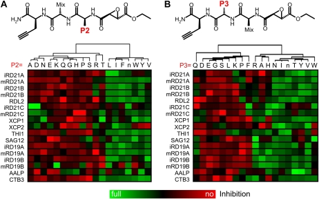
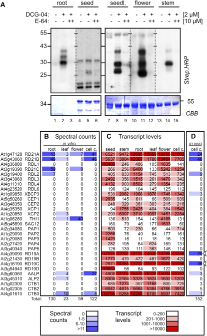
Papain-like cysteine proteases (PLCPs) are a large class of proteolytic enzymes associated with development, immunity, and senescence. Although many properties have been described for individual proteases, the distribution of these characteristics has not been studied collectively. Here, we analyzed 723 plant PLCPs and classify them into nine subfamilies that are present throughout the plant kingdom. Analysis of these subfamilies revealed previously unreported distinct subfamily-specific functional and structural characteristics. For example, the NPIR and KDEL localization signals are distinctive for subfamilies, and the carboxyl-terminal granulin domain occurs in two PLCP subfamilies, in which some individual members probably evolved by deletion of the granulin domains. We also discovered a conserved double cysteine in the catalytic site of SAG12-like proteases and two subfamily-specific disulfides in RD19A-like proteases. Protease activity profiling of representatives of the PLCP subfamilies using novel fluorescent probes revealed striking polymorphic labeling profiles and remarkably distinct pH dependency. Competition assays with peptide-epoxide scanning libraries revealed common and unique inhibitory fingerprints. Finally, we expand the detection of PLCPs by identifying common and organ-specific protease activities and identify previously undetected proteases upon labeling with cell-penetrating probes in vivo. This study provides the plant protease research community with tools for further functional annotation of plant PLCPs.


Figures

Figure 1.
Figure 1.
Phylogenetic subclassification of plant PLCPs. A, The unrooted phylogenetic tree of 723 plant PLCPs is subdivided into nine PLCP subfamilies (1–9). Arabidopsis PLCPs are indicated in the first column in color, and other studied PLCPs are indicated in the second column in black. Asterisks indicate that a crystal structure is available. Type members for each subfamily are shown in larger font. Key bootstrap values are indicated. The annotated phylogenetic tree with readable entries is available as Supplemental Figure S1. B, Distribution of PLCPs over subfamilies for plant species with more than 20 sequenced PLCPs. C, Nomenclature and subclassification of Arabidopsis PLCPs. The ATG accession codes of genes encoding putative PLCPs are followed by given names. The domain structure consists of a signal peptide (sp), prodomain (pro), protease domain with catalytic Cys (c), and in some cases a Pro-rich domain (p) and a granulin domain. The PLCPs studied biochemically in this work are marked in the right column. [See online article for color version of this figure.]
Figure 2.
Figure 2.
Conserved functional motifs in PLCP subfamilies. A, Positions and frequencies of functional motifs in each PLCP subfamily. SP, N-terminal signal peptide, predicted by SignalP; NPIR, vacuolar targeting signal at the N terminus of the prodomain; ERFNIN, structural motif in the prodomain (ExxxRxxxFxxNxxx{I/V}xxxN; allows one mismatch); Triad, the catalytic triad: Cys-His-Asn; KDEL, C-terminal retrieval signal for localization to the ER ({K/H}DEL); Granulin, C-terminal granulin domain containing the Cys pattern Cx5Cx5CCCx7Cx4CCx6CCx5CCx6Cx6C. B, Presence of the granulin domain in PLCP subfamilies 1 and 4. Granulin-containing proteases are indicated with black or red lines, and proteases lacking a granulin domain are indicated in gray or pink. Shown are only the trees of subfamilies 1 and 4 from Figure 1A. C, The phylogenetic tree of the granulin domain branches into the same subfamilies as the proprotease tree. Note that the subfamily 4 PLCPs (red; XBCP3-like) are grouped together. D, Conserved structural features of the granulin domain. Consensus sequences of the granulin domain of subfamilies 1 and 4 are aligned with those of RD21A and hGrnA. Disulfide bridging has been determined for hGrnA and is shown at the top. E, Illustration of the structure of the granulin domain of RD21A, modeled on hGrnA (2jye). The numbering of the disulfide bridge residues corresponds to that in D.
Figure 3.
Figure 3.
Conserved structural features in PLCP subfamilies. A, Positions of disulfide bridges and PGSs mapped onto the crystal structure of papain (1ppp) and cathepsin B (CathB; 3k9m). Cartoon models show the enzymes from the side with the α-helix domain (left) and β-sheet domain (right) and the catalytic triad (dotted spheres) with the catalytic Cys (cyan sticks) on top. The color code is as follows: α-helix (cyan); β-sheet (purple); loop (pink); extendable loop (blue); disulfide bridge (red); PGS (green). Numbers 1 to 9 correspond to conserved putative disulfide brides, summarized in B and C. B, Summary of the positions of putative disulfide bridges and PGSs in the mature protease domain. Positions are indicated for catalytic residues C, H, and N (black dashed lines), PGSs (NxS/T; green lines), and putative disulfide bridges (red lines). C, Frequency of conserved putative disulfide bridges, a double catalytic Cys (CCW), and PGS (NxS/T) in the different subfamilies. [See online article for color version of this figure.]
Figure 4.
Figure 4.
Protease activity profiling of representative PLCPs. A, Structures of MV201 and MV202. The E-64-based inhibitor group (red) contains an epoxide reactive group and a dipeptide carrying Leu (P2) and Tyr (P3) and is linked to the BODIPY fluorescent group (yellow) and either an azide minitag (green) or biotin (blue). B, PLCPs react with MV201. PLCPs were overexpressed in N. benthamiana by agroinfiltration in the presence of p19 silencing inhibitors. Extracts of agroinfiltrated leaves were labeled with 2 μm MV201 at pH 6 for 1 h, and labeled protein was detected from protein gels using fluorescence scanning. Asterisks indicate that, for identification purposes, proteins were labeled with MV202 and identified by in-gel digestion with trypsin and MS. This gel is a representative of at least three independent labeling experiments. [See online article for color version of this figure.]
Figure 5.
Figure 5.
pH-dependent labeling of PLCPs. N. benthamiana leaves overexpressing different PLCPs were labeled with 2 μm MV201 for 1 h at different pH levels. Fluorescent signals were quantified from protein gels by fluorescence scanning and plotted against pH. Each pH series was repeated at least once with similar outcome. [See online article for color version of this figure.]
Figure 6.
Figure 6.
PLCPs have distinct inhibitor sensitivities. Inhibitory fingerprinting is shown using P2 (A) and P3 (B) scanning epoxide libraries. These libraries contain a fixed amino acid at the P2 or P3 position, respectively, and an isokinetic mixture of 19 amino acids at the P3 or P2 position, respectively (top). Extracts containing different PLCPs were preincubated with a 10 μm epoxide library for 30 min and then labeled with 2 μm MV201 for 2 h. Labeled proteins were quantified from fluorescent gels and normalized relative to the median signal. The data were quantified and clustered based on the similarity of inhibition profiles. Similar inhibition data were obtained with repetition experiments.
Figure 7.
Figure 7.
PLCP activity and expression in different organs and in vivo. A, Activity profiles of PLCPs of different organs. Crude extracts of various organs were labeled with 2 μm DCG-04, either with or without a 30-min pretreatment with E-64 (10 μm), for 2.5 h at pH 6. Biotinylated proteins were detected on protein blots using streptavidin-horseradish peroxidase. CBB, Coomassie Brilliant Blue. B, Spectral counts of labeled PLCPs detected in different organs. Extracts from various organs were labeled with MV202 at pH 6, and labeled proteins were purified on avidin beads, separated on protein gels, excised, digested with trypsin, and analyzed by LC-MS/MS. The spectrum of peptides with greater than 95% confidence was counted for each PLCP; these are summarized in Supplemental Table S1. C, Transcript levels of all PLCPs in different organs. These data were extracted from Genevestigator (Hruz et al., 2008). D, In vivo labeling of PLCPs reveals activities of RD19A and RD19B. Arabidopsis cell cultures were labeled with 5 μm MV201. Proteins were extracted and coupled to Bio≡ using click chemistry. Biotinylated proteins were purified on avidin beads, separated on denaturing acrylamide gels, excised, digested with trypsin, and analyzed by LC-MS/MS. The spectrum of peptides with greater than 95% confidence was counted for each PLCP. [See online article for color version of this figure.]

References

    1. Ahmed SU, Rojo E, Kovaleva V, Venkataraman S, Dombrowski JE, Matsuoka K, Raikhel NV. (2000) The plant vacuolar sorting receptor AtELP is involved in transport of NH(2)-terminal propeptide-containing vacuolar proteins in Arabidopsis thaliana. J Cell Biol 149: 1335–1344 - PMC - PubMed
    1. Altschul SF, Gish W, Miller W, Myers EW, Lipman DJ. (1990) Basic local alignment search tool. J Mol Biol 215: 403–410 - PubMed
    1. Avci U, Petzold HE, Ismail IO, Beers EP, Haigler CH. (2008) Cysteine proteases XCP1 and XCP2 aid micro-autolysis within the intact central vacuole during xylogenesis in Arabidopsis roots. Plant J 56: 303–315 - PubMed
    1. Baldauf SL. (2003) Phylogeny for the faint of heart: a tutorial. Trends Genet 19: 345–351 - PubMed
    1. Bateman A, Bennett HPJ. (2009) The granulin gene family: from cancer to dementia. Bioessays 31: 1245–1254 - PubMed

Publication types

MeSH terms

LinkOut - more resources