. 2012 Mar 13;109(11):4221-6.
doi: 10.1073/pnas.1117019109. Epub 2012 Feb 27.

RNase H-mediated degradation of toxic RNA in myotonic dystrophy type 1

Affiliations

RNase H-mediated degradation of toxic RNA in myotonic dystrophy type 1

Johanna E Lee et al. Proc Natl Acad Sci U S A. .

Abstract

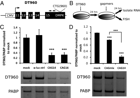
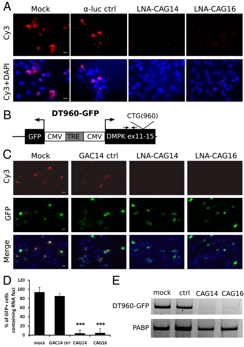
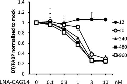
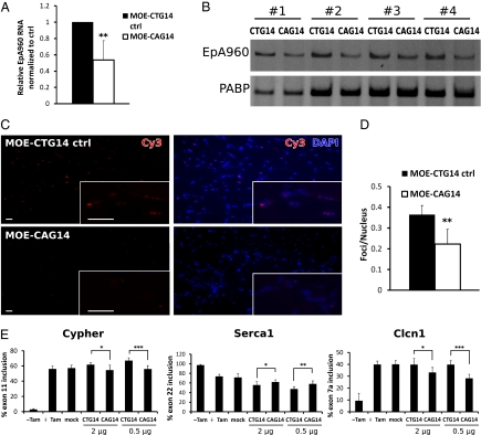
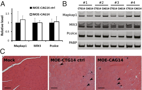
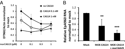
Myotonic dystrophy type 1 (DM1) is an RNA-dominant disease caused by abnormal transcripts containing expanded CUG repeats. The CUG transcripts aggregate in the nucleus to form RNA foci and lead to nuclear depletion of Muscleblind-like 1 (MBNL1) and stabilized expression of CUGBP Elav like family 1 (CELF1), both of which are splicing regulatory proteins. The imbalance of these proteins results in misregulation of alternative splicing and neuromuscular abnormalities. Here, we report the use of antisense oligonucleotides (ASOs) as a therapeutic approach to target the pathogenic RNA in DM1. We designed chimeric ASOs, termed gapmers, containing modified nucleic acid residues to induce RNase H-mediated degradation of CUG-repeat transcripts. The gapmers selectively knockdown expanded CUG transcripts and are sufficient to disrupt RNA foci both in cell culture and mouse models for DM1. Furthermore, combination of gapmers with morpholino ASOs that help release binding of MBNL1 to the toxic RNA can potentially enhance the knockdown effect. Additional optimization will be required for systemic delivery; however, our study provides an alternative strategy for the use of ASOs in DM1 therapy.


Conflict of interest statement

Conflict of interest statement: C.F.B. is an employee of Isis Pharmaceuticals, Inc., and may materially benefit either directly or indirectly through stock options.

Figures

Fig. 1.
Fig. 1.
RNase H-mediated degradation of expanded DMPK transcript in cell culture. (A) Diagram of the DT960 minigene construct. The DT960 minigene contains the human DMPK genomic segment with exons 11–15 and 960 interrupted CTG repeats expressed by a CMV promoter/enhancer. Primer pairs for RT-PCR [E15upF (forward) and E15upR (reverse)] are located in exon 15 upstream of the repeats (indicated by arrows). (B) Schematic of the experimental strategy. DT960 plasmid was transiently transfected into COSM6 cells, followed by transfection of gapmers 24 h later. RNA isolation or fluorescence in situ hybridization (FISH) was performed the next day. (C) (Upper) Standard RT-PCR showed 70% reduction of DT960 RNA in cells treated with 50 nM of LNA-CAG14 or LNA-CAG16 gapmers. A gapmer complementary to luciferase (α-luc) served as control. Poly-A binding protein (PABP) was used as internal control. The results of four independent transfection experiments were averaged (***P < 0.001). (Lower) Representative gel image used for quantification. (D) (Upper) Standard RT-PCR results. MOE-CAGmix treatment resulted in 25% decrease of the DT960 RNA, whereas the MOE-CAG14 gapmer induced 80% reduction compared with mock. The data represent the average of three independent transfection experiments (***P < 0.001). (Lower) Representative gel image used for quantification.
Fig. 2.
Fig. 2.
Disruption of RNA foci by CAG gapmers in cell culture. (A) RNA foci containing DT960 RNA were detected by FISH using Cy3-labeled probes. Nuclei were counterstained with DAPI (Lower). All images were taken at the same exposure time. (Scale bars: 20 μm.) (B) Diagram of the DT960-GFP construct. The expression of DMPK RNA and GFP are controlled by a bidirectional tetracycline responsive element (TRE), which is activated by the transactivator in the presence of doxycycline (TetON). Primers (E15upF and E15upR) for RT-PCR are indicated with arrows. (C) FISH was performed on cells transfected with transfection reagent only (mock), GAC14 control gapmer, LNA-CAG14 and LNA-CAG16. All images were taken at the same exposure time. (Scale bars: 20 μm.) (D) Bar graph represents average percent of GFP+ cells containing RNA foci. For the results, ≥7 microscopic fields and a total of >90 cells were counted from three independent transfection experiments. (***P < 0.001). (E) Reduction of DT960 transcript was confirmed by standard RT-PCR. Poly-A binding protein (PABP) was used as internal control.
Fig. 3.
Fig. 3.
CAG gapmers preferentially degrade RNAs containing expanded CUG repeats. Cells expressing identical DMPK transcripts except containing 12, 40, 240, 480, or 960 CUG repeats were treated with increased dosage (0, 0.1, 0.3, 1, 3, 10 nM) of LNA-CAG14 gapmer. LNA-CAG14 had no effect on RNA containing 12 CUG repeats. DMPK transcripts containing longer repeats are affected at lower concentrations. Data represent average of three independent experiments.
Fig. 4.
Fig. 4.
CAG gapmer administration reduces expanded CUG RNA levels and aberrant splicing in a DM1 mouse model. (A) Real-time RT-PCR indicates a 50% decrease of the relative EpA960 transcript level in the TA muscle 2 wk after administration of MOE-CAG14 compared with the MOE-CTG14 control (n = 6 mice, triplicate assays per sample, **P < 0.01). β-actin was used as the internal control. (B) Standard RT-PCR shows a decrease in EpA960 transcript in muscle treated with MOE-CAG14 compared with the MOE-CTG14 control. Representative results from four treated mice (numbered) are shown. (C) Fewer RNA foci were detected in muscle treated with MOE-CAG14 gapmer compared MOE-CTG14 control. Nuclei were stained using DAPI (Right). All images were taken at the same exposure time. (Inset) Higher magnification of foci. (Scale bars: 20 μm.) (D) Quantification reveals 40% reduction of the number of foci per nucleus in MOE-CAG14 muscle compared with control. Five microscopic fields were counted per muscle. Bar graph represents the average of [number of foci/number of nuclei] each field (n = 5 mice). (E) RT-PCR quantification of inclusion of alternative exons from three misregulated splicing events in DM1. Lanes are as follows: EpA960/HSA-Cre mice not injected with tamoxifen, n = 3 (-Tam); untreated mice 2 wk post tam, n = 4 (+Tam); mock-treated mice 2 wk post tam, n = 4 (mock); tamoxifen-induced EpA960/HSA-Cre mouse muscle injected with MOE-CTG14 control (2 μg), n = 6 ; MOE-CAG14 (2 μg), n = 6; MOE-CTG14 control (0.5 μg), n = 7; MOE-CAG14 (0.5 μg), n = 7 (*P < 0.05, **P < 0.01, ***P < 0.001).
Fig. 5.
Fig. 5.
Secondary effects of CAG gapmers. (A) Real-time RT-PCR of three gene transcripts containing ≥8 CUG repeats. RNA levels are expressed as arbitrary units relative to β-actin, then normalized to the mean expression of control muscle. (n = 4 mice, triplicate assays per sample, P > 0.5 for each). (B) No consistent difference in expression level was seen by standard RT-PCR. Data from four mice is shown. (C) Hematoxylin and eosin staining of TA muscle cross-sections. Central nuclei (arrowheads) and regions with multiple nuclei (asterisks) are present in muscle treated with MOE-CTG14 control and MOE-CAG14 gapmers. (Scale bar = 100 μm.)
Fig. 6.
Fig. 6.
Combined effect of CAG gapmers and morpholinos. (A) Following expression of DT960 RNA in COSM6 cells, LNA-CAG14 gapmer (0, 0.1, 1 nM) was transfected into COSM6 cells with increasing doses (0, 0.1, 0.3, 1 μM) of CAG morpholino containing 13 nucleotides (morCAG13). β-actin was used as internal control. (B) Real-time RT-PCR revealed enhanced decrease of EpA960 transcript level when MOE-CAG14 (2 μg) was combined with morCAG25 (20 μg). EpA960 transcript levels from PBS-treated muscle served as control (mock). n = 4–6 mice per group, triplicates were done per sample. β-actin was used as internal control. (**P < 0.01, ***P < 0.001).

References

    1. Harper P.S. Myotonic Dystrophy. London: W.B. Saunders; 2001.
    1. Brook JD, et al. Molecular basis of myotonic dystrophy: expansion of a trinucleotide (CTG) repeat at the 3′ end of a transcript encoding a protein kinase family member. Cell. 1992;68:799–808. - PubMed
    1. Davis BM, McCurrach ME, Taneja KL, Singer RH, Housman DE. Expansion of a CUG trinucleotide repeat in the 3′ untranslated region of myotonic dystrophy protein kinase transcripts results in nuclear retention of transcripts. Proc Natl Acad Sci USA. 1997;94:7388–7393. - PMC - PubMed
    1. Miller JW, et al. Recruitment of human muscleblind proteins to (CUG)(n) expansions associated with myotonic dystrophy. EMBO J. 2000;19:4439–4448. - PMC - PubMed
    1. Kuyumcu-Martinez NM, Wang GS, Cooper TA. Increased steady-state levels of CUGBP1 in myotonic dystrophy 1 are due to PKC-mediated hyperphosphorylation. Mol Cell. 2007;28:68–78. - PMC - PubMed

Publication types

MeSH terms

LinkOut - more resources