. 2012 Mar 13;109(11):4239-44.
doi: 10.1073/pnas.1114981109. Epub 2012 Feb 27.

Systematic identification of type I and type II interferon-induced antiviral factors

Affiliations

Systematic identification of type I and type II interferon-induced antiviral factors

Su-Yang Liu et al. Proc Natl Acad Sci U S A. .

Abstract

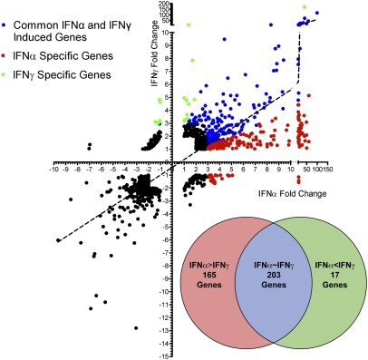
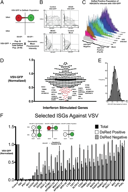
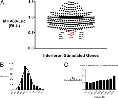
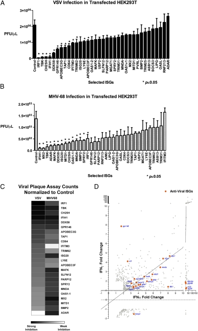
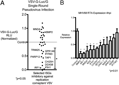
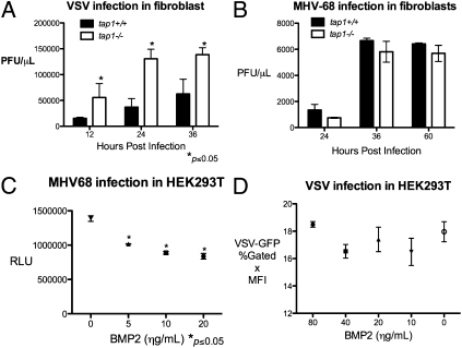
Type I and type II interferons (IFNs) are cytokines that establish the cellular antiviral state through the induction of IFN-stimulated genes (ISGs). We sought to understand the basis of the antiviral activity induced by type I and II IFNs in relation to the functions of their ISGs. Based on gene expression studies, we systematically identified antiviral ISGs by performing blinded, functional screens on 288 type I and type II ISGs. We assessed and validated the antiviral activity of these ISGs against an RNA virus, vesicular stomatitis virus (VSV), and a DNA virus, murine gammaherpes virus (MHV-68). Overall, we identified 34 ISGs that elicited an antiviral effect on the replication of either one or both viruses. Fourteen ISGs have uncharacterized antiviral functions. We further defined ISGs that affect critical life-cycle processes in expression of VSV protein and MHV-68 immediate-early genes. Two previously undescribed antiviral ISGs, TAP1 and BMP2, were further validated. TAP1-deficient fibroblasts were more susceptible to VSV infection but less so to MHV-68 infection. On the other hand, exogenous BMP2 inhibits MHV-68 lytic growth but did not affect VSV growth. These results delineate common and distinct sets of type I and type II IFN-induced genes as well as identify unique ISGs that have either broad or specific antiviral effects on these viruses.


Conflict of interest statement

The authors declare no conflict of interest.

Figures

Fig. 1.
Fig. 1.
Gene expression profile of BMMs treated for 2.5 h with IFNα and IFNγ at 62 U/mL and 1 U/mL, respectively. Axes represent fold change in response to IFNα or IFNγ over untreated cells. IFNα- and IFNγ-specific and commonly induced genes were categorized (see text) and are represented in a Venn diagram (Inset).
Fig. 2.
Fig. 2.
(A) Schematic of FACS-based assay to identify ISGs against VSV-GFP. HEK293T cells were transfected with individual ISGs with DsRed plasmid at a 3:1 ratio. DsRed-positive cells indicate cells that highly express the ISG. VSV growth (VSV-GFP) was calculated by the product of the percent GFP-positive cells and the geometric mean fluorescence intensity (MFI) of GFP in the DsRed-positive cells. (B) Contour maps of VSV-GFP in HEK293T cells transfected with the indicated ISGs and DsRed. (C) VSV-GFP was measured in the DsRed-positive population of selected ISGs transfected in HEK293T cells. TBK-1 was used as a positive control. (D) All 288 ISGs were screened by a FACS-based method. Each dot represents an ISG, and its effect on VSV-GFP expression is normalized to VSV-GFP in control transfected cells. ISGs that inhibited VSV-GFP expression over 50% are labeled and indicated in red. Values represent mean of duplicates. (E) Histogram of all ISGs and their effect on VSV-GFP normalized to VSV-GFP in control transfected cells. (F) Effect of individual ISGs on VSV-GFP expression in total population, DsRed-positive population, and DsRed-negative population was calculated. Values represent mean ± SEM from biological triplicates.
Fig. 3.
Fig. 3.
(A) Individual ISGs expressed in HEK293T cells and infected with MHV-68 at 0.25 MOI in quadruplicates. Cells were lysed at 9 hpi and luciferase activity was measured. Mean relative luminescence unit (RLU) are presented. (B) Histogram of all ISGs, and their effects on MHV-68-Luc were normalized to MHV-Luc in control transfected cells. (C) Selected ISGs from the screen were verified in two experiments in quadruplicate and normalized to control. Values are expressed as mean ± SEM.
Fig. 4.
Fig. 4.
(A and B) The inhibitory effects of 34 ISGs selected from previous screens against VSV (A) and MHV-68 (B) were measured by plaque assay. Values represent mean ± SEM. Data represent 1 of 3 experiments. Asterisks indicate significant difference compared with control by Student's t test (P < 0.05). (C) Heat map showing the inhibitory effect of selected ISGs on VSV and MHV-68 based on plaque assay. (D) Antiviral ISGs (orange) was graphed with respect to their fold induction by IFNα and IFNγ in microarray analyses (Fig. 1).
Fig. 5.
Fig. 5.
(A) ISGs that were most inhibitory against VSV in Fig. 4A were transfected in HEK293T cells for 36 h in triplicate and infected with VSVΔG-Luc reporter within a VSV-G envelope. Cells were lysed 10 hpi and luciferase activity was measured. BMP2 and MNDA, which did not inhibit VSV replication, were used as negative controls. Mean RLU are presented; data are representative of two independent experiments. (B) Eleven ISGs that were most inhibitory against MHV-68 were expressed in HEK293T cells for 36 h and infected with 0.2 MOI of MHV-68. Expression of the immediate-early gene RTA was measured by quantitative PCR at 4 hpi. IFITM3, which has no inhibitory effect on MHV-68, was used as a negative control. Data are representative of three independent experiments.
Fig. 6.
Fig. 6.
(A) tap1+/+ and tap1−/− tail-derived fibroblasts were infected with VSV at 0.1 MOI for the indicated time, and the supernatants were titered by plaque assay. Values represent mean ± SEM. (B) tap1+/+ and tap1−/− fibroblasts were infected the MHV-68 at 0.25 MOI, and the supernatants were collected at the indicated times and titered by plaque assay. Values represent mean ± SEM. (C) HEK293T were treated with hBMP2 at the indicated concentration for 12 h and infected with MHV-68-Luc at 0.25 MOI. Luciferase activity in the cell lysates was quantified at 9 hpi. Values represent mean ± SEM. RLU, relative luminescence unit. (D) HEK293T were treated with BMP2 at increasing concentrations for 12 h and infected with VSV at 0.01. VSV-GFP expression was measured by FACS at 9 hpi. Values represent mean ± SEM. MFI, geometric mean of fluorescence index.

References

    1. van den Broek MF, Müller U, Huang S, Zinkernagel RM, Aguet M. Immune defence in mice lacking type I and/or type II interferon receptors. Immunol Rev. 1995;148(1):5–18. - PubMed
    1. Sadler AJ, Williams BRG. Interferon-inducible antiviral effectors. Nat Rev Immunol. 2008;8:559–568. - PMC - PubMed
    1. Schroder K, Hertzog PJ, Ravasi T, Hume DA. Interferon-γ: An overview of signals, mechanisms and functions. J Leukoc Biol. 2004;75(2):163–189. - PubMed
    1. Schoggins JW, et al. A diverse range of gene products are effectors of the type I interferon antiviral response. Nature. 2011;472:481–485. - PMC - PubMed
    1. Flaño E, Kim I-J, Woodland DL, Blackman MA. γ-Herpesvirus latency is preferentially maintained in splenic germinal center and memory B cells. J Exp Med. 2002;196:1363–1372. - PMC - PubMed

Publication types

MeSH terms

Substances

Associated data