. 2012 Apr 4;31(7):1752-63.
doi: 10.1038/emboj.2012.25. Epub 2012 Feb 28.

miR-493 induction during carcinogenesis blocks metastatic settlement of colon cancer cells in liver

Affiliations

miR-493 induction during carcinogenesis blocks metastatic settlement of colon cancer cells in liver

Koji Okamoto et al. EMBO J. .

Abstract

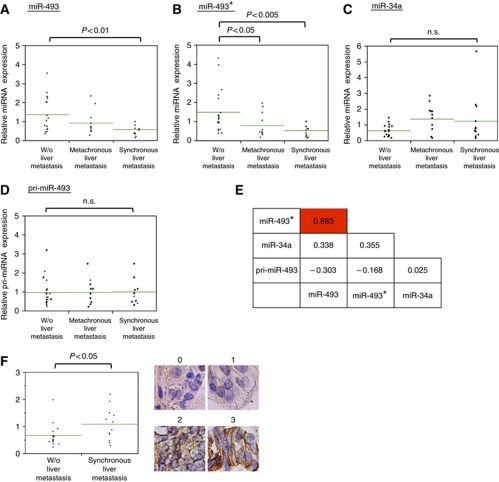
Liver metastasis is a major lethal complication associated with colon cancer, and post-intravasation steps of the metastasis are important for its clinical intervention. In order to identify inhibitory microRNAs (miRNAs) for these steps, we performed 'dropout' screens of a miRNA library in a mouse model of liver metastasis. Functional analyses showed that miR-493 and to a lesser extent miR-493(*) were capable of inhibiting liver metastasis. miR-493 inhibited retention of metastasized cells in liver parenchyma and induced their cell death. IGF1R was identified as a direct target of miR-493, and its inhibition partially phenocopied the anti-metastatic effects. High levels of miR-493 and miR-493(*), but not pri-miR-493, in primary colon cancer were inversely related to the presence of liver metastasis, and attributed to an increase of miR-493 expression during carcinogenesis. We propose that, in a subset of colon cancer, upregulation of miR-493 during carcinogenesis prevents liver metastasis via the induction of cell death of metastasized cells.


Conflict of interest statement

The authors declare that they have no conflict of interest.

Figures

Figure 1
Figure 1
Functional screening for miRNAs that inhibit liver metastasis of colon cancer cells. (A) Schematic for functional screening of miRNAs that inhibit liver metastasis of colon cancer cells. (1) HCT116 cells that were infected with a human miRNA lentivirus library were injected into the spleens of NOG mice. (2, 3) Two weeks after the injection, library-introduced cells were recovered from the spleen and liver, and used to isolate genomic DNA. (4, 5) The introduced miRNA was amplified from the genomic DNA by PCR, labelled with Cy3 (spleen) or Cy5 (liver), and the Cy5/Cy3 ratio of each library-introduced miRNA was determined with the custom-made microarray (see Materials and methods). (6) miRNAs with low Cy5/Cy3 values were regarded as positive ‘dropout’ clones. (B) Bright-phase (left) and GFP (right) images of metastasized liver (2 weeks after splenic injection). (C) Bright-phase (left) and GFP (right) images of the metastasized liver at higher magnification. Arrow shows cut surface. (D) HE section of the metastasized liver (2 weeks after splenic injection). (E) Immunostaining of the metastasized liver (5 days after splenic injection). The metastasized liver was immunostained with E-cadherin (left and lower right), or stained with HE (upper right). The right panels show magnified images. Arrow indicates a metastasized HCT116 focus. (F) A list of ‘dropout’ clones with low Cy5/Cy3 ratio (>10-fold). miRNAs with low P-values (P<0.01) are shown in bold. P-values were calculated by standardizing each of the four experimental data sets by Z-score transformation and then by examining null hypothesis that the dropout value of certain miRNA is identical to the average of dropout values of all miRNAs in the library. Welch’s t-test was performed to calculate P-value.
Figure 2
Figure 2
miR-493 expression inhibits liver metastasis of colon cancer cells. (A) Schematic for evaluation of inhibitory effects of each miRNA on liver metastasis. HCT116/GFP cells introduced with individual miRNA were mixed with HCT116/RFP cells at a 1:1 ratio, and the mixture was used for splenic injection to generate liver metastasis. (B, C) Inhibition of liver metastasis by miRNA precursors. HCT116/GFP cells were infected with the lentiviruses that express the indicated miRNA precursors, and the mixtures of the infected HCT116/GFP cells and HCT116/RFP cells were used for the splenic injection. (B) Metastasized cells were isolated from the liver 14 days after the splenic injection, and a number of GFP-positive and RFP-positive cells were counted by flow cytometry. The inhibitory effect of each miRNA was evaluated by calculating GFP/RFP ratio. (C) Inhibition of liver metastasis by miRNAs (day 14). GFP/RFP dual fluorescence images for the indicated miRNA precursors were taken. (D) miR-493 expression in colon cancer cells and colon epithelial cells.miR-493 and miR-493* expression of the indicated cells was determined by RT–qPCR. (E) RT–qPCR analyses of miR-493 (left) and miR-493* (right) in FHC cells and HCT116 cells infected with the lentiviruses that express the miR-493 precursor. (F) Inhibition of liver metastasis by miR-493 and miR-493* mimics. GFP/RFP dual fluorescence images for the indicated miRNAs were taken as described in (C). (G) Inhibition of liver metastasis by miR-493 and miR-493* mimics. Inhibitory effect of each miRNA was evaluated as described in (B). (H) Inhibitory effect of miR-493 and miR-493* mimics in DLD-1 cells. For liver metastasis assays, the inhibitory effect was evaluated by counting the number of GFP/RFP fluorescent foci 14 days after the splenic injection. For in vitro growth assays, the same mixture of cells was grown under normal culture condition for 14 days, and the GFP/RFP ratios were calculated by flow cytometry.
Figure 3
Figure 3
miR-493 expression induces cell death of colon cancer cells in metastasized liver. (A) GFP/RFP dual fluorescence images (higher magnification) of metastasized liver at day 2 or day 4 after splenic injection. HCT116/GFP cells were transfected with the indicated miRNA, and used for splenic injection as described in Figure 2C. (B) A number of GFP-positive and RFP-positive foci in (A) were counted and the relative ratio of GFP/RFP foci was calculated in each experiment. (C) Annexin V assays of metastasized cells transfected with miR-493. HCT116/RFP cells transfected with the indicated miRNA mimics were injected into the spleen. Three days after splenic injection, Alexa 488-Annexin V was injected via a tail vein, and the fraction of liver-metastasized HCT116/RFP cells that were positive for Annexin V staining was quantified under fluorescent microscope. Upper pictures show representative staining of HCT116/RFP cells with Alexa 488-Annexin V.
Figure 4
Figure 4
IGF1R is a direct target of miR-493 and partially mediates the inhibition of liver metastasis by miR-493. (A) Two-cycle Venn diagram shows the overlap of predicted target genes of miR-493 (40 genes) and genes inhibited by miR-493 expression (421 genes). (B) HCT116 cells were transfected with the indicated miRNA mimics (left panels), or infected with the control or the miR-493-expressing lentiviruses (right panels), and used for western blot analyses with the indicated antibodies. (C) Sequence comparison of IGF1R 3′UTR (positions 1311–1317), the mutated IGF1R 3′UTR, and human miR-493. (D) Transient luciferase assays. The indicated reporter plasmid was transfected into HCT116 cells together with control miRNA or miR-493 mimic, and luciferase activity was measured 2 days after transfection. (E) Western blot analyses in DLD-1 cells as shown in (B). (F) Western blot analyses in HCT116/GFP cells after transfection of control or the designated IGF1R siRNA. (G) Inhibition of liver metastasis by IGF1R siRNAs. The inhibitory effect of each siRNA was evaluated as described in Figure 2B. (H) Annexin V assays of metastasized HCT116 cells that were transfected with control or IGF1R siRNA. A fraction of Annexin V-positive cells after transfection of the indicated siRNA is shown as presented in Figure 3C.
Figure 5
Figure 5
A high level of miR-493 expression is associated with the absence of liver metastasis of colon cancer. (AD) RT–qPCR analyses of (A) miR-493, (B) miR-493*, (C) miR-34a, and (D) pri-miR-493 from the designated groups of primary colon tumours. Green lines indicate average values of each group. The relative miRNA expression level in each sample was normalized by the average of total samples. (E) Pearson's correlation coefficient between the designated variables. (F) Levels of IGF1R immunostaining of primary tumour without metastasis or with synchronous metastasis. Right panels indicate representative immunostaining from grade 0 to 3. Each immunostaining was quantified as described in Materials and methods.
Figure 6
Figure 6
miR-493 expression is induced during carcinogenesis in a subset of colon cancers. (A, C) RT–qPCR analyses of (A) miR-493 and (C) miR-493* in 27 primary tumours (14 cases without liver metastasis and 13 cases with synchronous liver metastasis) and the corresponding non-cancerous specimens. (B, D) Ratios of (B) miR-493 and (D) miR-493* levels between tumour and non-cancerous specimens analysed in (A) and (C), respectively. (E) Model for the role of miR-493 in suppressing liver metastasis in a subset of liver tumours.

Similar articles

Cited by

References

    1. Bartel DP (2009) MicroRNAs: target recognition and regulatory functions. Cell 136: 215–233 - PMC - PubMed
    1. Bouvet M, Tsuji K, Yang M, Jiang P, Moossa AR, Hoffman RM (2006) In vivo color-coded imaging of the interaction of colon cancer cells and splenocytes in the formation of liver metastases. Cancer Res 66: 11293–11297 - PubMed
    1. Chaffer CL, Weinberg RA (2011) A perspective on cancer cell metastasis. Science 331: 1559–1564 - PubMed
    1. Chambers AF, Groom AC, MacDonald IC (2002) Dissemination and growth of cancer cells in metastatic sites. Nat Rev Cancer 2: 563–572 - PubMed
    1. Chitnis MM, Yuen JS, Protheroe AS, Pollak M, Macaulay VM (2008) The type 1 insulin-like growth factor receptor pathway. Clin Cancer Res 14: 6364–6370 - PubMed

Publication types

Associated data