. 2012 Jul;40(12):5560-8.
doi: 10.1093/nar/gks179. Epub 2012 Feb 28.

Engineered zinc finger nickases induce homology-directed repair with reduced mutagenic effects

Affiliations

Engineered zinc finger nickases induce homology-directed repair with reduced mutagenic effects

Cherie L Ramirez et al. Nucleic Acids Res. 2012 Jul.

Abstract

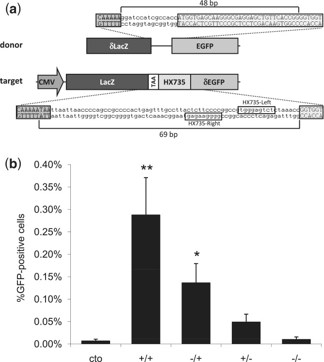
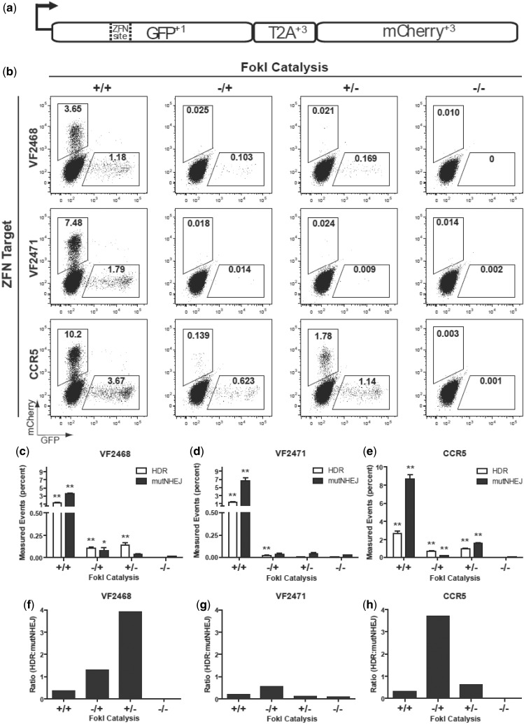
Engineered zinc finger nucleases (ZFNs) induce DNA double-strand breaks at specific recognition sequences and can promote efficient introduction of desired insertions, deletions or substitutions at or near the cut site via homology-directed repair (HDR) with a double- and/or single-stranded donor DNA template. However, mutagenic events caused by error-prone non-homologous end-joining (NHEJ)-mediated repair are introduced with equal or higher frequency at the nuclease cleavage site. Furthermore, unintended mutations can also result from NHEJ-mediated repair of off-target nuclease cleavage sites. Here, we describe a simple and general method for converting engineered ZFNs into zinc finger nickases (ZFNickases) by inactivating the catalytic activity of one monomer in a ZFN dimer. ZFNickases show robust strand-specific nicking activity in vitro. In addition, we demonstrate that ZFNickases can stimulate HDR at their nicking site in human cells, albeit at a lower frequency than by the ZFNs from which they were derived. Finally, we find that ZFNickases appear to induce greatly reduced levels of mutagenic NHEJ at their target nicking site. ZFNickases thus provide a promising means for inducing HDR-mediated gene modifications while reducing unwanted mutagenesis caused by error-prone NHEJ.


Figures

Figure 1.
Figure 1.
A qualitative in vitro assay to detect cleavage and nicking by ZFNs and ZFNickases. An asymmetrically positioned full ZFN target site is placed within a DNA fragment that has been labeled on both its 5′-ends with 6-FAM fluorescent dye (depicted in blue). Only 6-FAM labeled strands will be detected in the denaturing capillary electrophoresis assay. In the example shown, the ZFN target site is positioned toward the left end of the DNA fragment. In this configuration, if nicking of the top strand occurs, this results in the generation of one short and one full-length 6-FAM labeled product. If nicking of the bottom strand occurs, this results in the generation of one medium-length and one full-length 6-FAM labeled product. Cleavage of both strands results in the generation of short and medium-length products. Sample electropherograms are shown with arbitrary intensity units on the y-axis and DNA strand length on the x-axis. DNA strands expected from nicking or cleavage reactions are designated by black arrows. Note that full-length DNA strands due to incomplete enzyme reactions may be present in addition to the expected products.
Figure 2.
Figure 2.
Site-specific nicking of DNA in vitro by ZFNickases. Substrates labeled with 6-FAM fluorescent dye harboring (a) HX735, (b) VF2468, (c) VF2471 or (d) CCR5 binding sites were incubated with active Left/active Right (+/+), inactive Left/active Right (−/+), active Left/inactive Right (+/−) and inactive Left/inactive Right (−/−) ZFN monomers. Cleavage products were subjected to denaturing capillary electrophoresis. Axes are arbitrary intensity units (y-axis) and DNA strand length (x-axis). The y-axis is differentially scaled for each plot, whereas the x-axis is scaled uniformly for all plots. Representative electropherograms are shown, but all experiments were performed in triplicate (data not shown). Note that the HX735, VF2468 and VF2471 targets were cloned into the pCP5 vector that results in asymmetric placement left of center within the substrate similar to the configuration depicted in Figure 1. However, the CCR5 target is cloned into pBAC-lacZ, which results in binding site placement right of center relative to the substrate; when the top strand is cleaved in this configuration, the fragment generated is longer than when the bottom strand is cleaved.
Figure 3.
Figure 3.
Assessment of ZFNickase-mediated HDR using a human cell-based chromosomal EGFP reporter assay. (a) A schematic of the U2OS.LacZ-HX735-∂GFP reporter construct integrated in a U2OS cell line. Note that the orientation of the binding site in the reporter is inverted relative to the configuration at the HX735 endogenous locus, for which the Left and Right designations were originally (but arbitrarily) assigned. (b) ZFN and ZFNickase-mediated HDR in a U2OS EGFP reporter line. Cells were co-transfected with the donor plasmid and plasmids encoding HX735 ZFN pairs composed of active Left/active Right (+/+), inactive Left/active Right (−/+), active Left/inactive Right (+/−) and inactive Left/inactive Right (−/−) FokI domains. The graph shows the percentage of EGFP-positive cells 8 days following transfection. Statistically significant differences in HDR-based gene correction relative to donor-only control (cto) are indicated by * (P < 0.05) or ** (P < 0.01).
Figure 4.
Figure 4.
Assessment of ZFNickase-mediated HDR and NHEJ using a human cell-based TLR assay. (a) Schematic of the ‘TLR’ HDR-mediated correction of the EGFP gene with a co-transfected donor template results in EGFP-positive cells. Mutagenic NHEJ events at the nuclease target site result in mCherry-positive cells. (b) Representative flow cytometry plots showing percentages of EGFP-positive and mCherry-positive cells following transfection of TLR cell lines with plasmid encoding the indicated ZFNs and ZFNickases and the donor template. In the experiments shown, cells have been gated for BFP expression (encoded by the plasmid harboring the donor template) to normalize for transfection efficiencies. (c–e) Bar graphs showing mean percentages of EGFP-positive and mCherry-positive cells for experiments performed with the VF2468, VF2471 and CCR5 ZFNs and ZFNickases. Results were derived from three independent experiments with SEM shown. Statistically significant differences in HDR and mutagenic NHEJ rates relative to donor-only control (−/−) are indicated by * (P < 0.05) or ** (P < 0.01). (f–h) Ratios of percentage of EGFP-positive cells to percentage of mCherry-positive cells for the VF2468, VF2471 and CCR5 ZFNs and ZFNickases using the data from (c–e). Data used to create Figure 4c–h is available in Supplementary Table S3.

Comment in

References

    1. Kim YG, Cha J, Chandrasegaran S. Hybrid restriction enzymes: zinc finger fusions to Fok I cleavage domain. Proc. Natl Acad. Sci. USA. 1996;93:1156–1160. - PMC - PubMed
    1. Mani M, Smith J, Kandavelou K, Berg JM, Chandrasegaran S. Binding of two zinc finger nuclease monomers to two specific sites is required for effective double-strand DNA cleavage. Biochem. Biophys. Res. Commun. 2005;334:1191–1197. - PMC - PubMed
    1. Doyon Y, Vo TD, Mendel MC, Greenberg SG, Wang J, Xia DF, Miller JC, Urnov FD, Gregory PD, Holmes MC. Enhancing zinc-finger-nuclease activity with improved obligate heterodimeric architectures. Nat. Methods. 2011;8:74–79. - PubMed
    1. Miller JC, Holmes MC, Wang J, Guschin DY, Lee YL, Rupniewski I, Beausejour CM, Waite AJ, Wang NS, Kim KA, et al. An improved zinc-finger nuclease architecture for highly specific genome editing. Nat. Biotechnol. 2007;25:778–785. - PubMed
    1. Szczepek M, Brondani V, Buchel J, Serrano L, Segal DJ, Cathomen T. Structure-based redesign of the dimerization interface reduces the toxicity of zinc-finger nucleases. Nat. Biotechnol. 2007;25:786–793. - PubMed

Publication types

Substances