Review
. 2012:59:131-75.
doi: 10.1007/978-94-007-3015-1_5.

Role of phosphoinositides at the neuronal synapse

Affiliations
Review

Role of phosphoinositides at the neuronal synapse

Samuel G Frere et al. Subcell Biochem. 2012.

Abstract

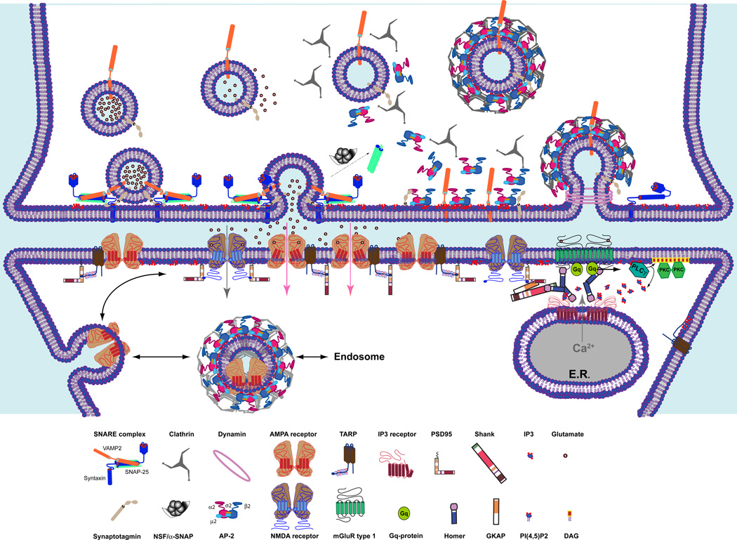
Synaptic transmission is amongst the most sophisticated and tightly controlled biological phenomena in higher eukaryotes. In the past few decades, tremendous progress has been made in our understanding of the molecular mechanisms underlying multiple facets of neurotransmission, both pre- and postsynaptically. Brought under the spotlight by pioneer studies in the areas of secretion and signal transduction, phosphoinositides and their metabolizing enzymes have been increasingly recognized as key protagonists in fundamental aspects of neurotransmission. Not surprisingly, dysregulation of phosphoinositide metabolism has also been implicated in synaptic malfunction associated with a variety of brain disorders. In the present chapter, we summarize current knowledge on the role of phosphoinositides at the neuronal synapse and highlight some of the outstanding questions in this research field.


Figures

Fig. 1
Fig. 1. Neurotransmission at glutamatergic synapses
Presynatically, synaptic vesicles (SVs) filled with glutamate are localized in proximity to the plasma membrane (PM) and are docked at the active zone after the assembling of the SNARE complex (and other proteins such as Munc18, not depicted here) composed of the PM-associated syntaxin-1, the cytosolic SNAP-25 and the SV membrane-associated synaptobrevin. Upon the arrival of a calcium (Ca2+) influx, the vesicular and the plasma membranes fused and the content of the SV is released in the synaptic cleft. Following SV collapse, the SNARE complex is disassembled by the ATPase N-ethylmaleimide-sensitive factor (NSF) and its adaptor α-SNAP. The fused vesicle is retrieved by CME, which starts by the recruitment of the clathrin adaptors, AP-2 to PM enriched in PtdIns(4,5)P2 and by its interaction with Syt-1. Clathrin molecules assemble into a lattice structure at the endocytic site and, along with a variety of tubulating factors, permit PM invagination and the formation of a CCP. Dynamin oligomers formed a helix that encircles the bud of the pit and triggers the fission and the individuation of a clathrin-coated vesicles. Quickly, the clathrin coat is removed and the AP-2-clathrin complex is disassembled. The SV is refilled with neurotransmitters and can undergo another cycle of exocytosis and endocytosis. Postsynaptically, released glutamate binds to the ionotropic AMPA and NMDA receptors and to the Gq-coupled metabotropic glutamate receptor. (mGluR) type 1 (the other types of mGluR are not depicted). Gating of the AMPA receptor by glutamate generates a net current influx that depolarizes the postsynaptic PM allowing the gating of glutamate-bound NMDA receptor and a slower influx of sodium and calcium. mGluR type 1 activation releases the Gq proteins and the hydrolysis of PtsIns(4,5)P2 by phospholipase Cγ into diacylglycerol (DAG) and inositol (1,4,5)-trisphosphate (IP3). DAG recruits to the PM and activates the protein kinase C, while IP3 binds to and opens the IP3 receptor (IP3 R), which liberates the calcium store in the endoplasmic reticulum (ER). In the figure, interactions of the receptors with the structural proteins that organized the postsynaptic density (PSD) is emphasized. NMDA binds directly to PSD-95 with a PDZ-binding motif localized at the C-terminal extremity of the receptor. AMPA receptor and mGluR type 1 are indirectly anchored to the PSD-95 complex via the transmembrane AMPA receptor regulatory proteins (TARP) and via Homer/Shank/GKAP, respectively. Homer also interacts with the IP3 R and allows the anchoring of the receptor in proximity to the mGluR type 1. Finally, AMPA receptors are mobile and move traffic between the synaptic and extrasynaptic zone, where they can be internalized by CME and be recycled via the endosome.
Fig. 2
Fig. 2. PtdIns(4,5)P2 role in endocytosis
The PtdIns4P-enriched membrane of the SV fused and merged with the PtdIns(4,5)P2-enriched PM liberating the neurotransmitter extracellularly. AP-2 is recruited by PtdIns(4,5)P2 with other accessory clathrin adaptors to the surface of the PM. The PM buds and invaginates to form a Ω-shaped pit covered by clathrin lattice and that comprised the SV proteins. Fission factor, dynamin gets recruited to the bud with endophilin and synaptojanin. Dynamin mediates the scission of its neck to release a free clathrin-coated vesicle (CCV), possibly facilitated by the hydrolysis of PtdIns(4,5)P2 by synaptojanin1 (Synj1). The PtdIns(4,5)P2 present on the CCV is dephosphorylated to PI(4)P by Synj1, thereby promoting the shading of the adaptors from the membrane and the uncoating reaction.

Similar articles

Cited by

References

    1. Adayev T, Chen-Hwang MC, Murakami N, Wang R, Hwang YW. MNB/DYRK1A phosphorylation regulates the interactions of synaptojanin 1 with endocytic accessory proteins. Biochem Biophys Res Commun. 2006;351:1060–1065. - PMC - PubMed
    1. Anderson RA, Boronenkov IV, Doughman SD, Kunz J, Loijens JC. Phosphatidylinositol phosphate kinases, a multifaceted family of signaling enzymes. J Biol Chem. 1999;274:9907–9910. - PubMed
    1. Ann K, Kowalchyk JA, Loyet KM, Martin TF. Novel Ca2+-binding protein (CAPS) related to UNC-31 required for Ca2+-activated exocytosis. J Biol Chem. 1997;272:19637–19640. - PubMed
    1. Aoyagi K, Sugaya T, Umeda M, Yamamoto S, Terakawa S, Takahashi M. The activation of exocytotic sites by the formation of phosphatidylinositol 4,5-bisphosphate microdomains at syntaxin clusters. J Biol Chem. 2005;280:17346–17352. - PubMed
    1. Arai Y, Ijuin T, Takenawa T, Becker LE, Takashima S. Excessive expression of synaptojanin in brains with Down syndrome. Brain Dev. 2002;24:67–72. - PubMed

Publication types

MeSH terms

LinkOut - more resources