

Onyx 18 embolisation of dural arteriovenous fistula via arterial and venous pathways: preliminary experience and evaluation of the short-term outcomes
- PMID: 22374275
- PMCID: PMC3587089
- DOI: 10.1259/bjr/25192972
Onyx 18 embolisation of dural arteriovenous fistula via arterial and venous pathways: preliminary experience and evaluation of the short-term outcomes
Abstract
Objective: This paper mainly focuses on our preliminary experience and short-term outcome evaluation of embolisation of non-cavernous dural arteriovenous fistulas (ncsDAVFs) and cavernous sinus dural arteriovenous fistulas (csDAVFs) using Onyx 18 (ev3, Plymouth, MN), and in combination with coils, via arterial and venous approaches, respectively.
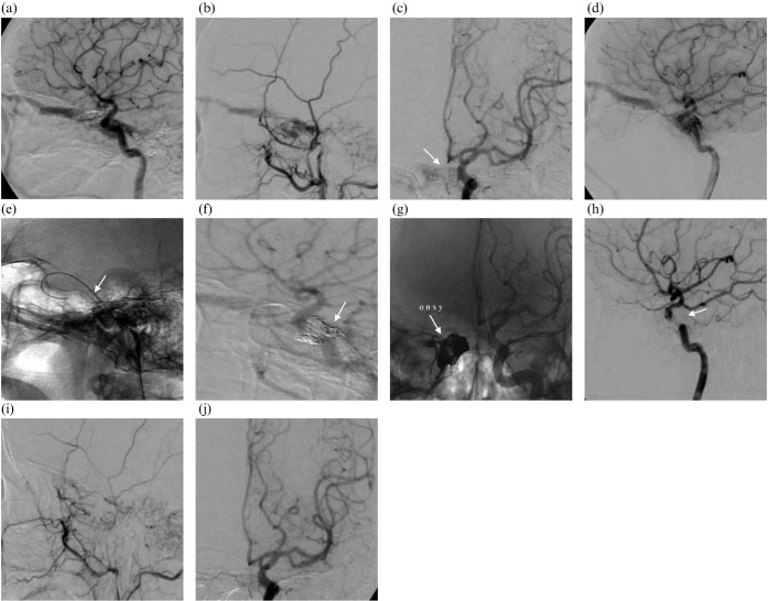
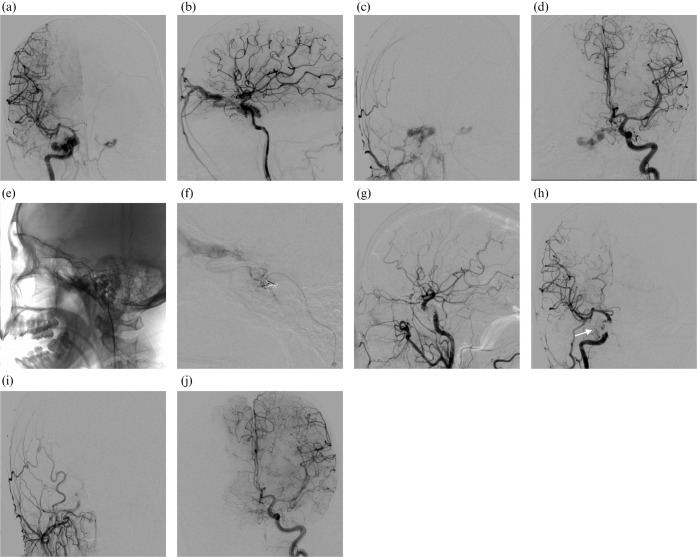
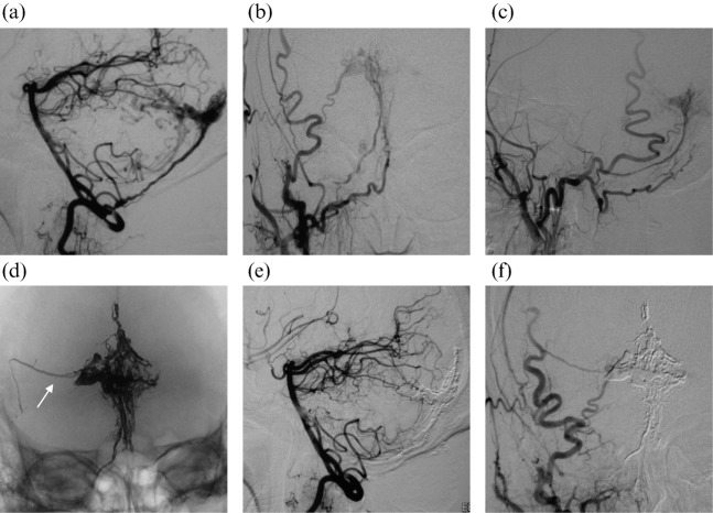
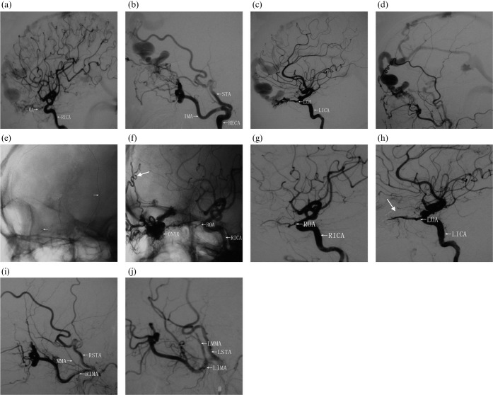
Methods: Between August 2008 and March 2010, 21 DAVFs (11 ncsDAVFs and 10 csDAVFs; age range: 28-68 years; 12 females and 9 males) were undertaken. Borden classification showed Type III in 1 and Type II in 10 ncsDAVFs, and Type II in 4 and Type I in 6 csDAVFs. Onyx 18 was used in 11 ncsDAVFs (10 via single feeder and 1 via 2 feeders). Onyx 18 or in combination with coils was used in 10 csDAVFs (9 via the inferior petrosal sinus and 1 via the superior ophthalmic vein).
Results: Total occlusion in immediate angiography was achieved in 18 cases (85.7%; 10 ncsDAVFs and 8 csDAVFs), and near-total occlusion in 1 ncsDAVF and 2 csDAVFs. Onyx 18 was migrated into normal vasculature in two ncsDAVFs without any sequelae. One csDAVF had VI cranial nerve palsy post-operatively, which completely recovered 2 weeks post-embolisation. Follow-up angiography at 3-12 months showed complete occlusion in 20 cases (95.2%; 10 ncsDAVFs and 10 csDAVFs). One ncsDAVF (4.8%) recurred after 3 months and was successfully re-embolised.
Conclusion: Preliminary results achieved after embolising 11 ncsDAVFs and 10 csDAVFs using Onyx 18 and in combination with coils via arterial and venous pathways, respectively, appeared to be safe, feasible and effective, as 95.2% of cases were totally occluded without any clinical sequelae.
Figures
Similar articles
-
Transvenous embolization with a combination of detachable coils and Onyx for a complicated cavernous dural arteriovenous fistula.Chin Med J (Engl). 2008 Sep 5;121(17):1651-5. Chin Med J (Engl). 2008. PMID: 19024093
-
Onyx as an adjunctive embolic material for transvenous embolization of cavernous sinus dural arteriovenous fistula after coiling.J Chin Med Assoc. 2025 Mar 1;88(3):261-266. doi: 10.1097/JCMA.0000000000001196. Epub 2024 Nov 25. J Chin Med Assoc. 2025. PMID: 39582118
-
Onyx embolization of dural arteriovenous fistulas of the cavernous sinus through the superior pharyngeal branch of the ascending pharyngeal artery.BMJ Case Rep. 2014 Apr 23;2014:bcr2013011067. doi: 10.1136/bcr-2013-011067. BMJ Case Rep. 2014. PMID: 24759156 Free PMC article.
-
Long-term follow-up of transarterial balloon-assisted Onyx embolization for endovascular treatment of dural arteriovenous fistulas: A single-institution case series and literature review.Clin Neurol Neurosurg. 2020 Dec;199:106256. doi: 10.1016/j.clineuro.2020.106256. Epub 2020 Oct 1. Clin Neurol Neurosurg. 2020. PMID: 33069089 Review.
-
Intra-arterial onyx embolisation of sphenobasilar sinus fistula using pressure cooker technique: case report and review of the literature.Neuroradiol J. 2021 Apr;34(2):131-134. doi: 10.1177/1971400920972512. Epub 2020 Nov 11. Neuroradiol J. 2021. PMID: 33176554 Free PMC article. Review.
Cited by
-
Angioarchitectural evolution of clival dural arteriovenous fistulas in two patients.Case Rep Ophthalmol. 2015 Mar 14;6(1):93-100. doi: 10.1159/000381176. eCollection 2015 Jan-Apr. Case Rep Ophthalmol. 2015. PMID: 25873894 Free PMC article.
-
Endovascular and surgical approaches of iatrogenic vertebrovertebral arteriovenous fistula.J Vasc Surg Cases Innov Tech. 2021 Feb 4;7(2):206-210. doi: 10.1016/j.jvscit.2020.12.019. eCollection 2021 Jun. J Vasc Surg Cases Innov Tech. 2021. PMID: 33997554 Free PMC article.
-
Focus on the target: Angiographic features of the fistulous point and prognosis of transvenous embolization of cavernous sinus dural arteriovenous fistula.Interv Neuroradiol. 2018 Apr;24(2):197-205. doi: 10.1177/1591019917751894. Epub 2018 Jan 19. Interv Neuroradiol. 2018. PMID: 29350092 Free PMC article.
References
-
- Newton TH, Cronqvist S. Involvement of dural arteries in intracranial arteriovenous malformations. Radiology 1969;93:1071–8 - PubMed
-
- Kiyosue H, Hori Y, Okahara M, Tanoue S, Sagara Y, Matsumoto S, et al. Treatment of intracranial dural arteriovenous fistulas: current strategies based on location and hemodynamics, and alternative techniques of transcatheter embolization. Radiographics 2004;24:1637–53 - PubMed
-
- Sarma D, ter Brugge K. Management of intracranial dural arteriovenous shunts in adults. Eur J Radiol 2003;46:206–20 - PubMed
MeSH terms
Substances
LinkOut - more resources
Full Text Sources
Medical
