Review
. 2012 Jun;85(1014):672-81.
doi: 10.1259/bjr/84443179. Epub 2012 Feb 28.

Review of the principal extra spinal pathologies causing sciatica and new MRI approaches

Affiliations
Review

Review of the principal extra spinal pathologies causing sciatica and new MRI approaches

A Ailianou et al. Br J Radiol. 2012 Jun.

Abstract

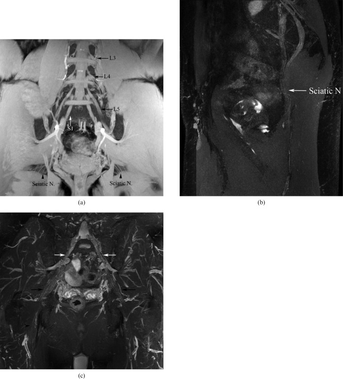
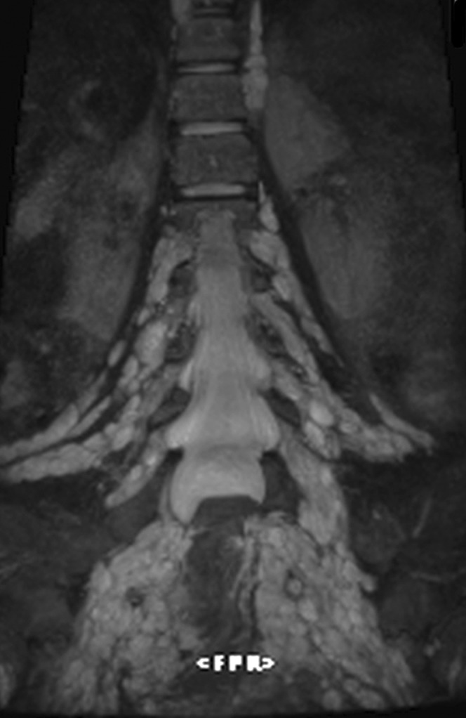
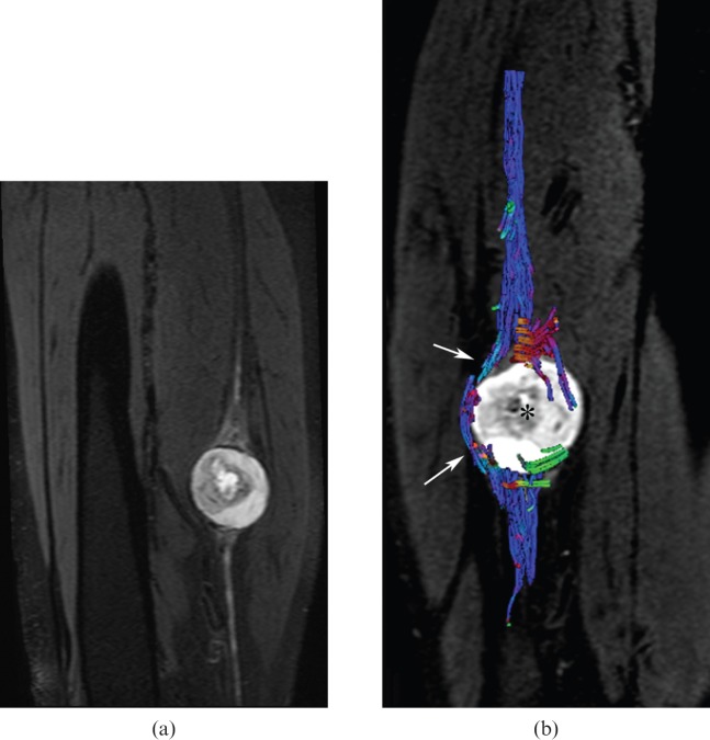
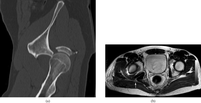
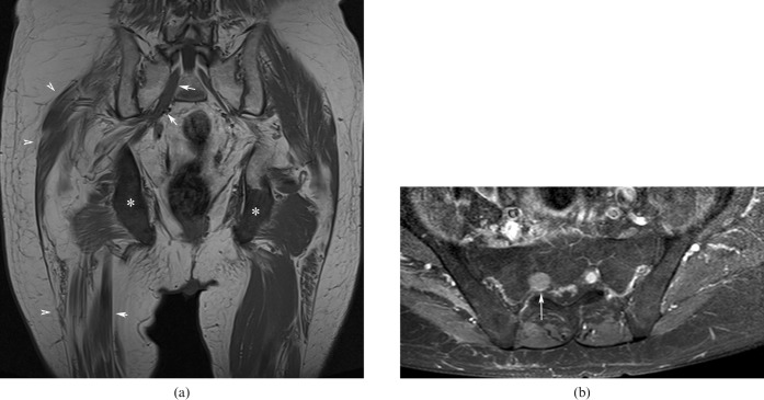
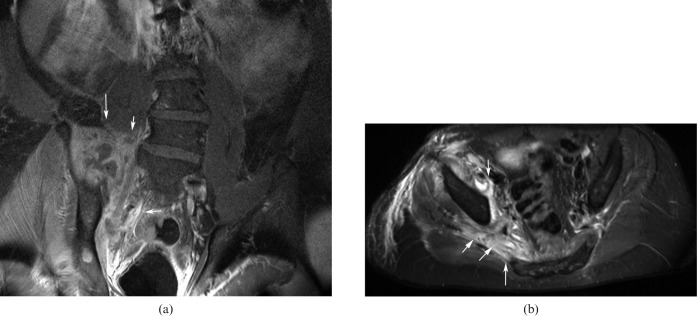
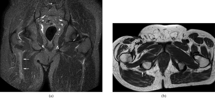
In this paper we illustrate the principal extraspinal pathologies causing sciatica and new approaches for the study of structures such as the lumbosacral plexus (LSP). Visualisation of the LSP in its entirety is difficult with conventional two-dimensional MRI sequences owing to its oblique orientation. In our institution, we have found that the utilisation of three-dimensional short tau inversion-recovery sampling perfection with application-optimised contrasts using different flip angle evolutions sequence is helpful, allowing multiplanar and maximum intensity projection reconstructions in the coronal oblique plane and curvilinear reformats through the plexus. Diffusion tensor imaging enables the observation of microstructural changes and can be useful in surgical planning. The normal anatomy of the LSP, its different extraspinal pathologies and differential diagnoses are thoroughly presented.


Figures

Figure 1
Figure 1
(a) Three-dimensional (3D) gradient echo T1 weighted image (echo time: 4.6 ms, repetition time: 8.6 ms) demonstrating the normal anatomy of the lumbosacral plexus (LSP). Note the L3–S1 nerves forming the LSP (arrows), the spinal ganglion (asterisk) and the sciatic nerves (arrowheads). (b) A curved reformatted 3D short tau inversion-recovery sampling perfection with application-optimised contrasts using different flip angle evolutions image (echo time: 3.1 ms, repetition time: 8.4 ms) demonstrates the sciatic nerve (arrow). (c) A coronal reformatted 3D short tau inversion-recovery multiplanar image (echo time: 123 ms, repetition time: 1800 ms) demonstrates the origin of the sciatic nerve (white arrows) and its flat configuration (black arrow), which more distally becomes tubular (arrowhead).
Figure 2
Figure 2
Contrast-enhanced T1 fat-saturated axial image (echo time: 15 ms, repetition time: 667 ms) showing a very well-delineated and enhanced ovoid mass corresponding to a neurofibroma (arrows) situated at the right sciatic nerve in a patient with neurofibromatosis Type 1.
Figure 3
Figure 3
Three-dimensional short tau inversion-recovery multiplanar (echo time: 150 ms, repetition time: 2000 ms) reformatted coronal image showing plexiform neurofibroma of both lumbosacral plexuses.
Figure 4
Figure 4
(a) Three-dimensional short tau inversion-recovery multiplanar reformatted sagittal image (echo time: 60 ms, repetition time: 6558 ms) and (b) tractography (echo time: 77 ms, repetition time: 9200 ms) revealing a schwannoma arising from the sciatic nerve (a) with neural tract displacement [arrows in (b)], but no infiltration by the neurogenic mass [asterisk in (b)].
Figure 5
Figure 5
Contrast-enhanced T1 fat-saturated coronal image (echo time: 10 ms, repetition time: 657 ms) demonstrating a cystic schwannoma originating from the right L3 nerve root (arrows).
Figure 6
Figure 6
(a) Unenhanced sagittally reconstructed CT image showing a posterior fracture dislocation of the hip joint. (b) T2 axial image after reduction shows the hyperintense and enlarged right sciatic nerve after hip reduction (arrow).
Figure 7
Figure 7
(a) Unenhanced T1 coronal image (echo time: 12 ms, repetition time: 400 ms) and (b) enhanced fat-saturated axial T1 image (echo time: 12 ms, repetition time: 400 ms), showing metastatic lesions in both ischial tuberosities [asterisks in (a)], atrophy of the glutei and proximal right thigh muscles [arrowheads in (a)] and secondary tumoral infiltration of the enlarged right sciatic nerve [arrows in (a, b)].
Figure 8
Figure 8
Contrast-enhanced T1 fat-saturated (a) coronal (echo time: 12 ms, repetition time: 400 ms) and (b) axial images illustrating a large abscess in the right psoas and glutei muscles in contact with the sciatic nerve (arrows).
Figure 9
Figure 9
(a) Short tau inversion-recovery coronal image (echo time: 60 ms, repetition time: 3685 ms) and (b) T2 spin echo axial image (echo time: 16 ms, repetition time: 943 ms) showing enlarged and hyperintense right sciatic nerve after amputation (arrows).

Similar articles

Cited by

References

    1. Yoshimoto M, Kawaguchi S, Takebayashi T, Isogai S, Kurata Y, Nonaka S, et al. Diagnostic features of sciatica without lumbar nerve root compression. J Spinal Disord Tech 2008;22:328–33 - PubMed
    1. Rohkmann R. Atlas de Poche de neurologie. Paris, France: Flammarion; 2003
    1. Vargas MI, Beaulieu J, Magistris MR, Della Santa D, Delavelle J. Clinical findings, electroneuromyography and MRI in trauma of the brachial plexus. J Neuroradiol 2007;34:236–42 - PubMed
    1. Chappell KE, Robson MD, Stonebridge-Foster A, Glover A, Allsop JM, Williams AD, et al. Magic angle effects in MR neurography. AJNR Am J Neuroradiol 2004;25:431–40 - PMC - PubMed
    1. Viallon M, Vargas MI, Jlassi H, Lövblad KO, Delavelle J. High-resolution and functional magnetic resonance imaging of the brachial plexus using an isotropic 3D T2 STIR (short term inversion recovery) SPACE sequence and diffusion tensor imaging. Eur Radiol 2008;18:1018–23 - PubMed