

320-detector row CT coronary angiography: effects of heart rate and heart rate variability on image quality, diagnostic accuracy and radiation exposure
- PMID: 22374285
- PMCID: PMC3587067
- DOI: 10.1259/bjr/92160185
320-detector row CT coronary angiography: effects of heart rate and heart rate variability on image quality, diagnostic accuracy and radiation exposure
Abstract
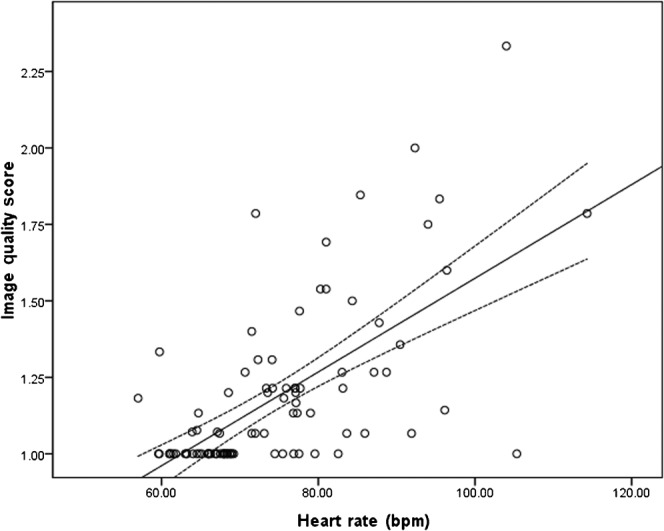
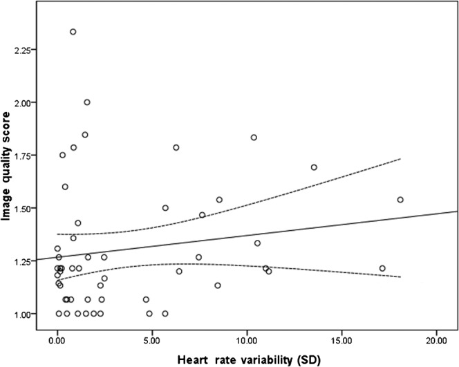
Objectives: To evaluate the effects of heart rate and heart rate variability on image quality, patient dose and diagnostic accuracy of 320-detector row CT.
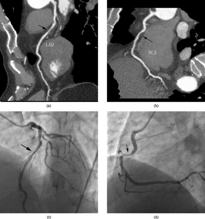
Methods: 94 patients were prospectively enrolled. Heart rate was defined as the mean value of different intervals elapsing between two consecutive R waves in an electrocardiogram (R-R intervals) and the heart rate variability was calculated as the standard deviation from the average heart rate. The image quality was evaluated by four grades, according to motion artefacts ("step artefacts" and "blurring artefacts"). The diagnostic accuracy was analysed in 43 patients who were scheduled for invasive coronary angiography (ICA). The coeffects of heart rate and heart rate variability on image quality, radiation dose and diagnostic accuracy were evaluated by multivariate regression.
Results: The mean image quality score was 1.2 ± 0.5 and the mean effective dose was 14.8 ± 9.8 mSv. The results showed that heart rate (74.0 ± 11.2 beats per minute) was the single factor influencing image quality (p<0.001) and radiation dose (p<0.001), while heart rate variability (3.7 ± 4.6) had no significant effect on them (p=0.16 and p=0.47, respectively). For 43 patients who underwent ICA, heart rate and heart rate variability showed no influence on the accuracy (p=0.17 and p=0.12, respectively). Overall sensitivity was 97.4% (37/38), specificity was 99.4% (351/353), positive predictive value was 94.9% (37/39) and negative predictive value was 99.7% (351/352).
Conclusion: 320-detector row CT, with improved longitudinal coverage of detector, resolves step artefact and high patient dose caused by irregular heart rate. However, it is still recommended to control heart rate to a lower level to eliminate blurring artefact and radiation dose.
Figures
References
-
- Pelberg RA, Mazur W, Clarke G, Szawaluk J. The what and why of cardiac CT angiography: data interpretation and clinical practice integration. Rev Cardiovasc Med 2009;10:152–63 - PubMed
-
- Brodoefel H, Burgstahler C, Tsiflikas I, Reimann A, Schroeder S, Claussen CD, et al. Dual-source CT: effect of heart rate, heart rate variability, and calcification on image quality and diagnostic accuracy. Radiology 2008;247:346–55 - PubMed
-
- Matt D, Scheffel H, Leschka S, Flohr TG, Marincek B, Kaufmann PA, et al. Dual-source CT coronary angiography: image quality, mean heart rate, and heart rate variability. AJR Am J Roentgenol 2007;189:567–73 - PubMed
-
- Weustink A, Neefjes L, Kyrzopoulos S, van Straten M, Neoh ER, Meijboom W, et al. Impact of heart rate frequency and variability on radiation exposure, image quality, and diagnostic performance in dual-source spiral CT coronary angiography. Radiology 2009;253:672–80 - PubMed
-
- Kitagawa K, Lardo A, Lima J, George R. Prospective ECG-gated 320 row detector computed tomography: implications for CT angiography and perfusion imaging. Int J Cardiovasc Imaging 2009;25:201–8
MeSH terms
LinkOut - more resources
Full Text Sources
Medical
Miscellaneous
