

Biglycan is a specific marker and an autocrine angiogenic factor of tumour endothelial cells
- PMID: 22374465
- PMCID: PMC3304426
- DOI: 10.1038/bjc.2012.59
Biglycan is a specific marker and an autocrine angiogenic factor of tumour endothelial cells
Abstract
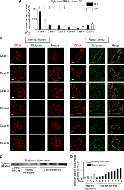
Background: We isolated tumour endothelial cells (TECs), demonstrated their abnormalities, compared gene expression profiles of TECs and normal endothelial cells (NECs) by microarray analysis and identified several genes upregulated in TECs. We focused on the gene encoding biglycan, a small leucine-rich repeat proteoglycan. No report is available on biglycan expression or function in TECs.
Methods: The NEC and TEC were isolated. We investigated the biglycan expression and function in TECs. Western blotting analysis of biglycan was performed on sera from cancer patients.
Results: Biglycan expression levels were higher in TECs than in NECs. Biglycan knockdown inhibited cell migration and caused morphological changes in TECs. Furthermore, immunostaining revealed strong biglycan expression in vivo in human tumour vessels, as in mouse TECs. Biglycan was detected in the sera of cancer patients but was hardly detected in those of healthy volunteers.
Conclusion: These findings suggested that biglycan is a novel TEC marker and a target for anti-angiogenic therapy.
Figures


Similar articles
-
Tumour endothelial cells in high metastatic tumours promote metastasis via epigenetic dysregulation of biglycan.Sci Rep. 2016 Jun 13;6:28039. doi: 10.1038/srep28039. Sci Rep. 2016. PMID: 27295191 Free PMC article.
-
CXCR7: a novel tumor endothelial marker in renal cell carcinoma.Pathol Int. 2012 May;62(5):309-17. doi: 10.1111/j.1440-1827.2012.02792.x. Epub 2012 Feb 21. Pathol Int. 2012. PMID: 22524658
-
CXCL12-CXCR7 axis is important for tumor endothelial cell angiogenic property.Int J Cancer. 2015 Dec 15;137(12):2825-36. doi: 10.1002/ijc.29655. Epub 2015 Jul 2. Int J Cancer. 2015. PMID: 26100110
-
Heterogeneity of tumor endothelial cells.Cancer Sci. 2013 Nov;104(11):1391-5. doi: 10.1111/cas.12251. Epub 2013 Sep 12. Cancer Sci. 2013. PMID: 23930621 Free PMC article. Review.
-
Tumour endothelial cells acquire drug resistance in a tumour microenvironment.J Biochem. 2013 Mar;153(3):243-9. doi: 10.1093/jb/mvs152. Epub 2013 Jan 3. J Biochem. 2013. PMID: 23293323 Review.
Cited by
-
The Extracellular Small Leucine-Rich Proteoglycan Biglycan Is a Key Player in Gastric Cancer Aggressiveness.Cancers (Basel). 2021 Mar 16;13(6):1330. doi: 10.3390/cancers13061330. Cancers (Basel). 2021. PMID: 33809543 Free PMC article.
-
Oncosuppressive functions of decorin.Mol Cell Oncol. 2015 Feb 25;2(3):e975645. doi: 10.4161/23723556.2014.975645. eCollection 2015 Jul-Sep. Mol Cell Oncol. 2015. PMID: 27308453 Free PMC article. Review.
-
Adsorption of cellular proteins to polyelectrolyte-functionalized gold nanorods: a mechanism for nanoparticle regulation of cell phenotype?PLoS One. 2014 Feb 6;9(2):e86670. doi: 10.1371/journal.pone.0086670. eCollection 2014. PLoS One. 2014. PMID: 24516536 Free PMC article.
-
Suprabasin as a novel tumor endothelial cell marker.Cancer Sci. 2014 Dec;105(12):1533-40. doi: 10.1111/cas.12549. Epub 2014 Nov 5. Cancer Sci. 2014. PMID: 25283635 Free PMC article.
-
Proteoglycans and their functions in esophageal squamous cell carcinoma.World J Clin Oncol. 2021 Jul 24;12(7):507-521. doi: 10.5306/wjco.v12.i7.507. World J Clin Oncol. 2021. PMID: 34367925 Free PMC article. Review.
References
-
- Akiyama K, Ohga N, Hida Y, Kawamoto T, Sadamoto Y, Ishikawa S, Maishi N, Akino T, Kondoh M, Matsuda A, Inoue N, Shindoh M, Hida K (2012) Tumor endothelial cells acquire drug resistance by MDR1 upregulation via VEGF signaling in tumor microenvironment. Am J Pathol 180(3): 1283–1293 - PubMed
-
- Bergers G, Benjamin LE (2003) Tumorigenesis and the angiogenic switch. Nat Rev Cancer 3: 401–410 - PubMed
-
- Bianco P, Fisher LW, Young MF, Termine JD, Robey PG (1990) Expression and localization of the two small proteoglycans biglycan and decorin in developing human skeletal and non-skeletal tissues. J Histochem Cytochem 38: 1549–1563 - PubMed
Publication types
MeSH terms
Substances
LinkOut - more resources
Full Text Sources
Miscellaneous
