. 2012 Oct;228(2):230-40.
doi: 10.1002/path.4011. Epub 2012 Jul 26.

Epigenetic disruption of cadherin-11 in human cancer metastasis

Affiliations
Free PMC article

Epigenetic disruption of cadherin-11 in human cancer metastasis

F Javier Carmona et al. J Pathol. 2012 Oct.
Free PMC article

Abstract

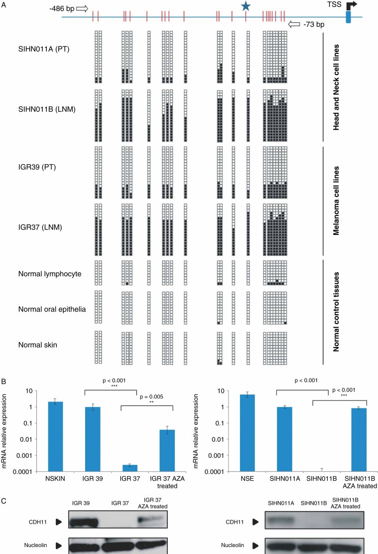
Little is known about the molecular events occurring in the metastases of human tumours. Epigenetic alterations are dynamic lesions that change over the natural course of the disease, and so they might play a role in the biology of cancer cells that have departed from the primary tumour. Herein, we have adopted an epigenomic approach to identify some of these changes. Using a DNA methylation microarray platform to compare paired primary tumour and lymph node metastatic cell lines from the same patient, we observed cadherin-11 promoter CpG island hypermethylation as a likely target of the process. We found that CDH11 DNA methylation-associated transcriptional silencing occurred in the corresponding lymph node metastases of melanoma and head and neck cancer cells but not in the primary tumours. Using in vitro and in vivo cellular and mouse models for depleted or enhanced CDH11 activity, we also demonstrated that CDH11 acts as an inhibitor of tumour growth, motility and dissemination. Most importantly, the study of CDH11 5'-CpG island hypermethylation in primary tumours and lymph node metastases of cancer patients showed this epigenetic alteration to be significantly confined to the disseminated cells. Overall, these results indicate the existence of metastasis-specific epigenetic events that might contribute to the progression of the disease.


Figures

Figure 1
Figure 1
Unsupervised clustering of the DNA methylation profiles obtained from the analyses of the 1505 CpG sites in the same patients' paired primary tumour/metastasis cells in melanoma (IGR39/IGR37) and head and neck cancer (SIHN011A/SIHN011B)
Figure 2
Figure 2
DNA methylation-associated transcriptional silencing of CDH11. (A) Bisulphite genomic sequencing of CDH11 promoter CpG Island. CpG dinucleotides are represented as short vertical lines and the transcriptional start site (TSS) is represented as a long black arrow over a blue stripe. The locations of the bisulphite genomic sequencing primers are indicated by white arrows. A minimum of eight single clones are shown for each sample. Presence of a methylated or unmethylated cytosine is indicated by a black or a white square, respectively. (B) Quantification of CDH11 mRNA expression levels in IGR39, IGR37, SIHN011A and SIHN011B cell lines and normal control tissues by qRT–PCR. Values are expressed as mean ± SD of three independent experiments, each conducted in triplicate; p values were obtained from Student's t-tests. (C) Quantification of CDH11 protein by western blot; nucleolin was used as the loading control
Figure 3
Figure 3
Restoration of CDH11 expression in metastatic cancer cells reduces growth, motility and dissemination. (A) Full-length cDNA sequence of CDH11 was cloned in pEGFP–IRES plasmid for stable transfection of cadherin-11 in SIHN011B cells. Two transfected clones (SIHN011B #1 and SIHN011B #2) are shown. (B) Cell proliferation differences between CDH11-expressing clones and the corresponding controls determined by the MTT assay and monitored for 6 days; values displayed were obtained from independent experiments with 15 replicates and expressed as mean ± SD; p values were obtained from Student's t-tests. (C) Colony formation assay. The number of colonies formed 2 weeks after seeding 1000 cells on 35 mm plates and maintained on selection media were quantified and plotted; p values were obtained from Student's t-tests. (D) Invasiveness assessment, using the Matrigel-coated Boyden chamber assay. The number of cells invading through the membrane was quantified after 48 h of incubation. Images show a representative field of the membrane, and were taken at × 20 magnification; values are expressed as mean ± SD; p values were obtained from Student's t-tests. (E) Inhibitory effects of CDH11 expression on tumourigenicity. Head and neck metastatic cells transfected with CDH11 or empty vector were injected into the flanks of SCID mice. Tumour weight and volume were measured after 21 days. Bars represent the standard error of the mean (SEM); p values were obtained from Mann–Whitney U-tests. The photographs show representative cases on day 21 after implantation. (F) The restoration of CDH11 expression in SIHN011B-transfected cells reduce their capacity to generate lymph node metastasis (detected by methylene blue, right) following injection in the mice tongue submucosa. (G) Metastasis assay through tail vein injection of tumour cells and analysis by H&E staining. Cancer cells with CDH11 expression (SIHN011B #1 and #2) showed a significant number of metastatic foci; p values were obtained from Mann–Whitney U-tests. The photographs show multiple metastatic nodules (black arrows) in SIHN011B-mock tail vein-injected mice
Figure 4
Figure 4
Depletion of CDH11 expression enhances growth and motility of cancer cells derived from primary tumours. (A) Depletion of CDH11 expression was achieved through stable CDH11 shRNA vector transfection (SIHN011A-57/5), compared with the scrambled shRNA-transfected control cells (SIHN011A-Scb). (B) Proliferation rates assessed by the MTT assay were registered over 5 days. Values represented were obtained from independent experiments with 15 replicates and expressed as mean ± SD; p value obtained from Student's t-test. (C) Colony formation assay. The numbers of colonies formed after 2 weeks were quantified and plotted; a significant increase in the number of colonies was noted in the CDH11-knockdown cells; p value was obtained from Student's t-test. (D) Invasiveness assessment using the Matrigel-coated Boyden chamber assay; the number of cells invading through the membrane was quantified after 48 h of incubation. Cadherin-11-depleted cells SIHN011A-57/5 had significant increased invasive capacities with respect to the control SIHN011A-Scb cells. Values are expressed as mean ± SD; p value was obtained by Student's t-test. Images show a representative field of the membrane and were taken at × 20 magnification. (E) Enhancing effects of CDH11 down-regulation on tumourigenicity in athymic nude mice. The head and neck metastatic cells shRNA depleted for CDH11 and the scrambled shRNA control cells were injected into the flanks of SCID mice. Tumour weight and volume were measured after 21 days. Bars represent SEM; p values were obtained from Mann–Whitney U-tests. The photographs show representative cases on day 21 after implantation. (F) Metastasis assay through tail vein injection of tumour cells and analysis by H&E staining. Cancer cells with depleted CDH11 expression (SIHN011A-57/5) showed a significantly higher number of metastatic foci. The photographs show metastatic nodules (black arrows) in the lung from SIHN011A-57/5 tail vein-injected mice
Figure 5
Figure 5
Epigenetic inactivation of CDH11 occurs preferentially in the lymph node metastases of cancer patients. (A) Methylation-specific PCR was performed on bisulphite-treated DNA extracted from primary tumour and lymph node metastases of head and neck cancer and melanoma patients. The presence of a band under the U or M lanes indicates unmethylated or methylated sequences, respectively. Normal lymphocytes (NLs) and in vitro methylated DNA (IVD) are shown as positive controls for the unmethylated and methylated sequences, respectively. The significant enrichment of CDH11 hypermethylation in lymph node metastasis in comparison to primary tumours is represented in the pie diagrams below; p value was obtained from Fisher's exact test. (B) CDH11 methylation-specific PCR analysis of matched primary tumour/lymph node metastasis from seven patients shows the confinement of CDH11 hypermethylation in the disseminated samples. (C) Expression of the CDH11 protein by immunohistochemistry in paired primary tumour/metastasis samples of the four head and neck cancer patients studied in Figure 5B. The presence of CDH11 CpG island hypermethylation was associated with CDH11 loss, whereas an unmethylated CDH11 CpG island was linked to the presence of the CDH11 protein

References

    1. Jemal A, Siegel R, Xu J, et al. Cancer statistics, 2010. CA Cancer J Clin. 2010;60:277–300. - PubMed
    1. Fidler IJ. The pathogenesis of cancer metastasis: The ‘seed and soil’ hypothesis revisited. Nat Rev Cancer. 2003;3:453–458. - PubMed
    1. Eccles SA, Welch DR. Metastasis: recent discoveries and novel treatment strategies. Lancet. 2007;369:1742–1757. - PMC - PubMed
    1. Carmona FJ, Esteller M. IDIBELL cancer conference on metastasis and angiogenesis. Cancer Res. 2011;71:6097–7101. - PubMed
    1. Ma L, Teruya-Feldstein J, Weinberg RA. Tumour invasion and metastasis initiated by microRNA-10b in breast cancer. Nature. 2007;449:682–688. - PubMed

Publication types

MeSH terms