Comparative Study
. 2012 Mar;71(3):323-33.
doi: 10.1002/ana.22686. Epub 2012 Feb 28.

Neutrophil protease inhibition reduces neuromyelitis optica-immunoglobulin G-induced damage in mouse brain

Affiliations
Comparative Study

Neutrophil protease inhibition reduces neuromyelitis optica-immunoglobulin G-induced damage in mouse brain

Samira Saadoun et al. Ann Neurol. 2012 Mar.

Abstract

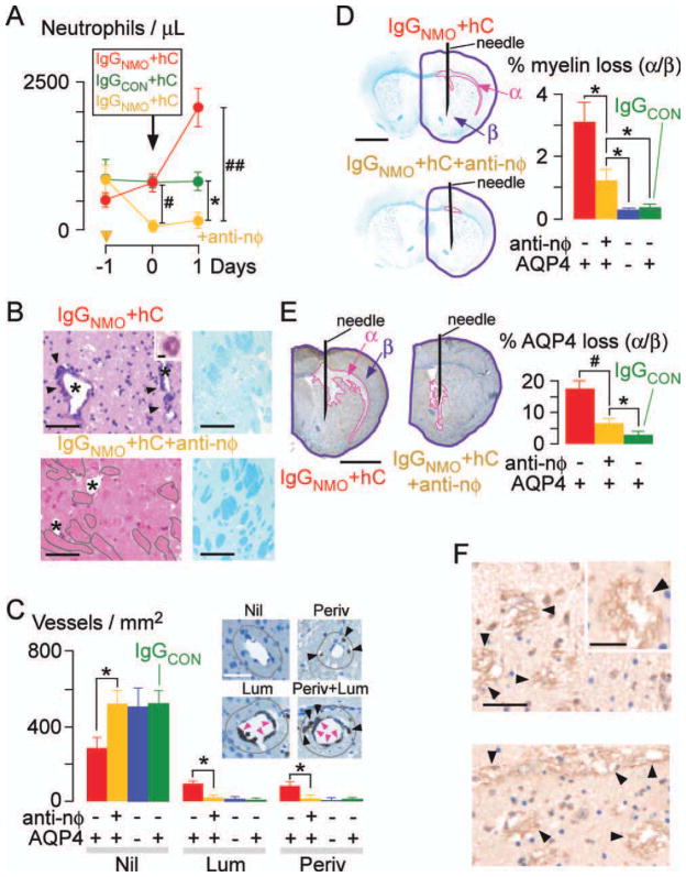
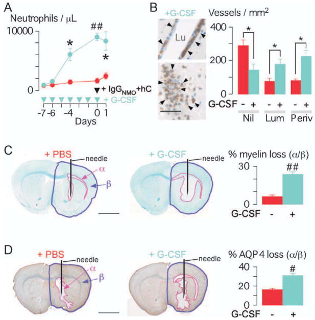
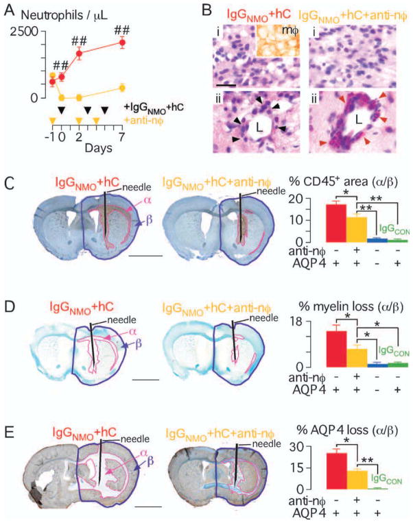
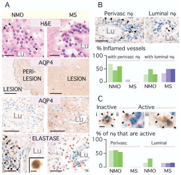
Objective: Neuromyelitis optica (NMO) is an inflammatory demyelinating disease of the central nervous system associated with pathogenic autoantibodies against the astrocyte water channel protein aquaporin-4 (AQP4). The presence of neutrophils is a characteristic feature in NMO lesions in humans. Neutrophils are not generally found in multiple sclerosis lesions. We evaluated the role of neutrophils in a mouse NMO model.

Methods: NMO lesions were produced in mice by intracerebral injection of immunoglobulin G (IgG) isolated from NMO patient serum and human complement. We previously reported that this mouse model produces the characteristic histological features of NMO, including perivascular complement activation, inflammatory cell infiltration, and loss of myelin, AQP4, and glial fibrillary acidic protein. Lesions are absent when AQP4 null mice are used or when IgG from non-NMO patients is injected.

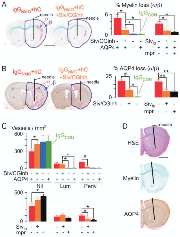
Results: We found remarkably reduced neuroinflammation, myelin loss, and AQP4 loss in brains of neutropenic mice at 24 hours and 7 days, and increased severity of NMO lesions in mice made neutrophilic by granulocyte colony stimulating factor. NMO lesions were greatly reduced by intracerebral administration of the neutrophil protease inhibitors Sivelestat and cathepsin G inhibitor I or by intraperitoneal injection of Sivelestat alone. Immunostaining of human NMO lesions for neutrophil elastase revealed many degranulating perivascular neutrophils, with no equivalent perivascular neutrophils in human multiple sclerosis lesions.

Interpretation: Our data implicate a central role of neutrophils in the pathogenesis of early NMO lesions and suggest the potential utility of neutrophil protease inhibitors such as Sivelestat in NMO therapy.


Conflict of interest statement

Potential Conflicts of Interest

A.S.V.: grants: EY13574 and DK35124 from the National Institutes of Health, and Guthy-Jackson Charitable Foundation. A.V.: grant: Oxford NIHR Biomedical Research Centre. M.C.P.: grant: Guthy-Jackson Charitable Foundation. P.W.: grant: Oxford NIHR Biomedical Research Centre; employment: University of Oxford; patents: patents for diagnostic assays for Lgi1 and Caspar2.

Figures

FIGURE 1
FIGURE 1
Neutropenia reduces brain damage at 24 hours after IgGNMO+ hC injection. (A) Peripheral neutrophil count. Antineutrophil (yellow) or isotype control (red and green) IgG was injected at day −1. Mice received IgGNMO+hC (red, yellow) or IgGCON+hC (green) at day 0 and were killed after 24 hours. (B) (Left) H&E: 0.5mm lateral to needle, non-neutropenic vs neutropenic (+anti-nφ) brain. (Inset) Perivascular neutrophil. * = lumen, arrowheads = perivascular inflammation, grey outline = intact white matter. (Right) LFB. (C) Neutrophil immunostain: Nil = no neutrophils, Lum = luminal neutrophils adherent to endothelium (pink arrowheads), Periv = perivascular neutrophils (black arrowheads) within 20μm radius (brown line), or Lum and Periv. Number of Nil, Lum, and Periv vessels/mm2. (D) (Left) LFB: Non-neutropenic vs neutropenic (+anti-nφ) brain. (Right) % loss of myelin (area-α/area-β). (E) (Left) AQP4 immunostain: non-neutropenic vs neutropenic (+anti-nφ) brain. (Right) % loss of AQP4 (area-α/area-β). (F) C5b-9 immunostain: 0.5mm lateral to needle, non-neutropenic vs neutropenic (+anti-nφ) brain. * = lumen, arrowheads = C5b-9. (Inset) C5b-9 (magnified). We injected 8 non-neutropenic (red), 8 neutropenic (yellow), and 5 non-neutropenic AQP4 null (blue) mice with IgGNMO+hC; 5 non-neutropenic mice received IgGCON+hC (green). Mean ± standard error. *p < 0.05, #p < 0.005, ##p < 0.0005 (vs IgGNMO+hC+anti-nφ). Bars = 2μm (B inset), 50μm (C inset, F inset), 100μm (B, F), 2mm (D, E).
FIGURE 2
FIGURE 2
Neutrophilia increases brain damage at 24 hours after IgGNMO + hC injection. (A) Peripheral neutrophil count. GCSF (blue) or PBS (red) was injected daily. All mice received IgGNMO+hC at day 0 and were killed after 24 hours. (B) Neutrophil immunostain (arrowheads): vessels from +G-CSF brain. Lu = lumen. Bottom picture: neutrophils occlude lumen. Number of Nil, Lum, and Periv vessels/mm2 in +PBS (red) vs +G-CSF (blue) mice. (C) (Left) LFB: +PBS vs +G-CSF brain. (Right) % loss of myelin (area-α/area-β). (D) (Left) AQP4 immunostain: +PBS vs +G-CSF brain. (Right) Loss of AQP4 (area-α/area-β). We used 4 +PBS vs 4 +G-CSF mice. Mean ± standard error. *p < 0.05, #p < 0.005, ##p < 0.0005 for +PBS vs +G-CSF. Bar = 50μm (B); 2mm (C, D).
FIGURE 3
FIGURE 3
Neutropenia reduces brain damage at 7 days after IgGNMO+ hC injection. (A) (Left) Peripheral neutrophil count. Antineutrophil (yellow) or isotype control (red) IgG was injected at days −1, 2, and 4. All mice received IgGNMO+hC at days 0, 3, and 5 and were killed on day 7. (B) H&E: non-neutropenic vs neutropenic (+anti-nφ) brain. (i) Brain parenchyma, inset = macrophage immunostain. (ii) Perivascular leukocytes, black arrowheads = mononuclear cells, red arrowheads = eosinophils, L = lumen. (C) (Left) CD45 immunostain: non-neutropenic vs neutropenic (+anti-nφ) brain. (Right) % inflammation (area-α/area-β). (D) (Left) LFB: non-neutropenic vs neutropenic (+anti-nφ) brain. (Right) % loss of myelin (area-α/area-β). (E) (Left) AQP4 immunostain: non-neutropenic vs neutropenic (+anti-nφ) brain. (Right) % loss of AQP4 (area-α/area-β). We injected 6 non-neutropenic (red), 6 neutropenic (yellow), and 5 non-neutropenic AQP4 null (blue) mice with IgGNMO+hC; 5 non-neutropenic mice received IgGCON+hC (green). Mean ± standard error. *p < 0.05, **p < 0.01, ##p < 0.0005 vs IgGNMO+hC+anti-nφ. Bar = 20 μm (B), 2 mm (C–E).
FIGURE 4
FIGURE 4
Neutrophil protease inhibitors reduce brain damage at 24 hours after IgGNMO+hC injection. (A) LFB, and (B) AQP4 immunostain. (Left) Brain injected with IgGNMO+hC alone (IgGNMO+hC) or with Sivelestat and cathepsin G inhibitor I (IgGNMO+hC+Siv/CGinh). (Right) % area-α/area-β (A) loss of myelin and (B) loss of AQP4 after IgGNMO+hC (6 mice, red), IgGNMO+hC+Siv/CGinh (6 mice, orange), IgGNMO+hC (5 AQP4 null mice, blue), or IgGCON+hC (5 mice, green). (Far Right) % area-α/area-β (A) loss of myelin and (B) loss of AQP4 after injecting IgGNMO+hC without (6 mice, red) vs with i.p. Sivelestat (6 mice, orange) vs with i.p. methylprednisolone (5 mice, black). (C) Number of Nil, Lum, and Periv vessels/mm2. (Top) IgGNMO+hC (6 mice, red), IgGNMO+hC+Siv/CGinh (6 mice, orange), IgGNMO+hC (5 AQP4 null mice, blue), or IgGCON+hC (5 mice, green). (Bottom) IgGNMO+hC without (6 mice, red) vs with i.p. Sivelestat (6 mice, orange) vs. with i.p. methylprednisolone (5 mice, black). (D) Brain at 24 hours after intracerebral injection of NE and Cath G: H&E (top), LFB (middle), AQP4 immunostain (bottom). Mean ± standard error. *p < 0.05, **p < 0.01, #p < 0.005 vs IgGNMO+hC+Siv/CGinh or +Sivip. Bar = 2mm (A, B, D).
FIGURE 5
FIGURE 5
Abundant neutrophil elastase within human NMO but not MS lesions. (A) NMO vs MS brain lesions. H&E: Arrows = neutrophils (black), eosinophils (blue), mononuclear cells (orange). AQP4 immunostain: (Top) Lesion and perilesion, (Bottom) Vessel in lesion. Lu = lumen. NE immunostain: Perivascular region. Arrows = NE+ (black) and NE (orange) leukocytes. (Inset) NE+ cell. (B) (Top) Vessel with perivascular (no intraluminal) and 1 with intraluminal (no perivascular) neutrophils. (Bottom) % inflamed vessels with perivascular NE+ cells and % with circulating (luminal) NE+ cells in 3 NMO (green) vs 3 MS (purple) samples. (C) Top (i) Inert and (ii,iii) active neutrophils. (i) NE (black arrows) is intracellular (red arrows). (ii) Extracellular NE. (iii) Elongated neutrophil (black arrows). (Bottom) Number of perivascular and luminal active neutrophils in 3 NMO (green) and 3 MS (purple) samples. Bar = 20μm (A: H&E), 0.5mm (A: AQP4 top), 100μm (A: AQP4 bottom), 50μm (A: Elastase, B), 5μm (A: Elastase inset, C).

References

    1. Wingerchuk DM, Lennon VA, Lucchinetti CF, et al. The spectrum of neuromyelitis optica. Lancet Neurol. 2007;6:805–815. - PubMed
    1. Jarius S, Paul F, Franciotta D, et al. Mechanisms of disease: aquaporin- 4 antibodies in neuromyelitis optica. Nat Clin Pract Neurol. 2008;4:202–214. - PubMed
    1. Lennon VA, Kryzer TJ, Pittock SJ, et al. IgG marker of optic-spinal multiple sclerosis binds to the aquaporin-4 water channel. J Exp Med. 2005;202:473–477. - PMC - PubMed
    1. Jarius S, Wildemann B. AQP4 antibodies in neuromyelitis optica: diagnostic and pathogenetic relevance. Nat Rev Neurol. 2010;6:383–392. - PubMed
    1. Lennon VA, Wingerchuk DM, Kryzer TJ, et al. A serum autoantibody marker of neuromyelitis optica: distinction from multiple sclerosis. Lancet. 2004;364:2106–2112. - PubMed

Publication types