. 2012 Apr;153(4):1925-35.
doi: 10.1210/en.2011-1900. Epub 2012 Feb 28.

Hematopoetic prostaglandin D synthase: an ESR1-dependent oviductal epithelial cell synthase

Affiliations

Hematopoetic prostaglandin D synthase: an ESR1-dependent oviductal epithelial cell synthase

Phillip J Bridges et al. Endocrinology. 2012 Apr.

Abstract

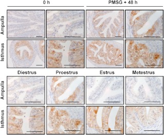
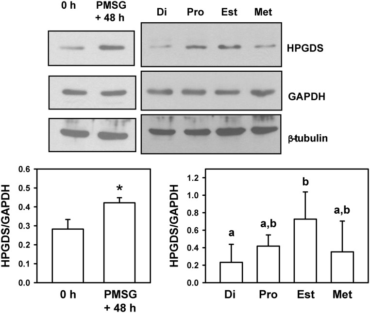
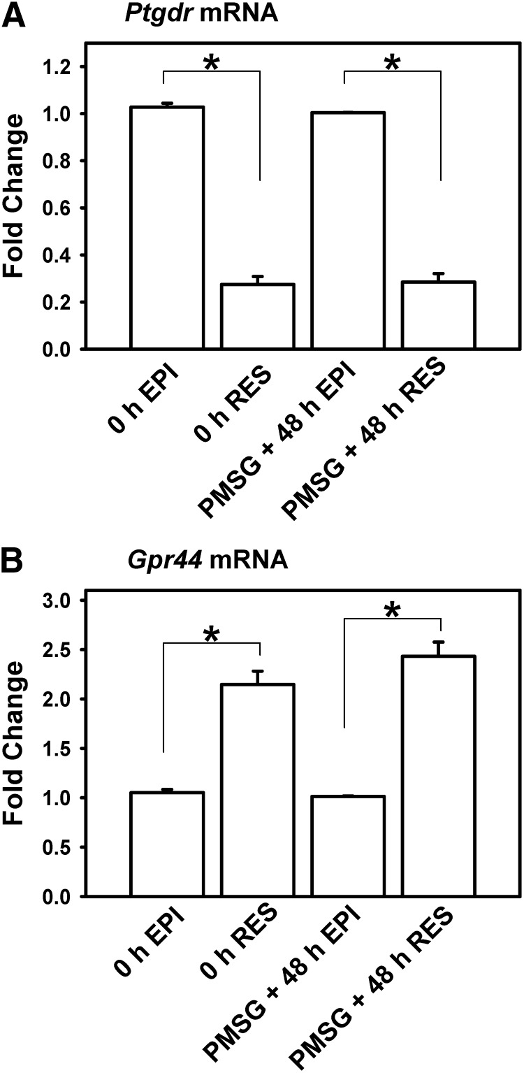
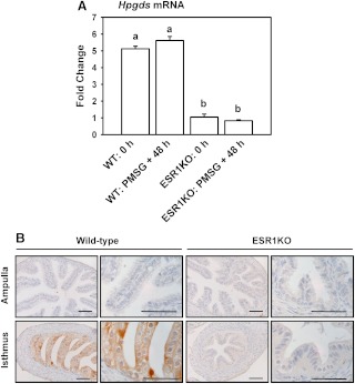
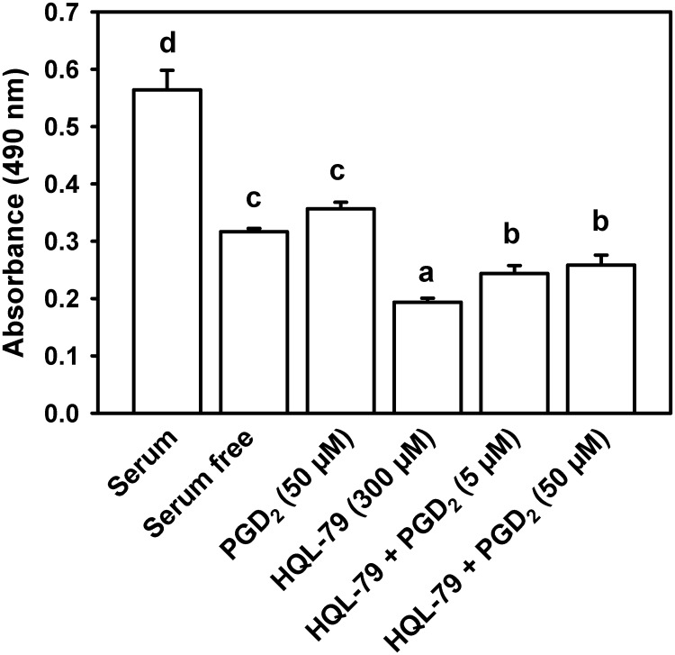
Oviductal disease is a primary cause of infertility, a problem that largely stems from excessive inflammation of this key reproductive organ. Our poor understanding of the mechanisms regulating oviductal inflammation restricts our ability to diagnose, treat, and/or prevent oviductal disease. Using mice, our objective was to determine the spatial localization, regulatory mechanism, and functional attributes of a hypothesized regulator of oviductal inflammation, the hematopoietic form of prostaglandin D synthase (HPGDS). Immunohistochemistry revealed specific localization of HPGDS to the oviduct's epithelium. In the isthmus, expression of HPGDS was consistent. In the ampulla, expression of HPGDS appeared dependent upon stage of the estrous cycle. HPGDS was expressed in the epithelium of immature and cycling mice but not in the oviducts of estrogen receptor α knockouts. Two receptor subtypes bind PGD₂: PGD₂ receptor and G protein-coupled receptor 44. Expression of mRNA for Ptgdr was higher in the epithelial cells (EPI) than in the stroma (P < 0.05), whereas mRNA for Gpr44 was higher in the stroma than epithelium (P < 0.05). Treatment of human oviductal EPI with HQL-79, an inhibitor of HPGDS, decreased cell viability (P < 0.05). Treatment of mice with HQL-79 increased mRNA for chemokine (C-C motif) ligands 3, 4, and 19; chemokine (C-X-C motif) ligands 11 and 12; IL-13 and IL-17B; and TNF receptor superfamily, member 1b (P < 0.02 for each mRNA). Overall, these results suggest that HPGDS may play a role in the regulation of inflammation and EPI health within the oviduct.


Figures

Fig. 1.
Fig. 1.
Immunohistochemical localization of HPGDS in the ampulla and isthmus of the mouse oviduct. Representative images are shown. Upper panels, Immature mice were killed at 23 d of age (0 h) or treated with 5 IU of PMSG to induce follicular development and killed 48 h later. Lower panel, Eight- to ten-week-old regularly cycling mice were killed on each day of the estrous cycle. Tissues were fixed in Bouin's fixative and embedded in paraffin. Scale bars, 50 μm.
Fig. 2.
Fig. 2.
Detection of HPGDS in whole oviducts of mice. Upper panels, Images of representative Western blottings are shown. Upper left, Immature mice were killed at 23 d of age (0 h) or treated with 5 IU of PMSG to induce follicular development and killed 48 h later. Upper right, Eight- to ten-week-old regularly cycling mice were killed on each day of the estrous cycle. Di, Diestrus; Pro, proestrus; Est, estrus; Met, metestrus. GAPDH and β-tubulin were used as loading controls. Lower panels, Quantification of Western blottings (HPGDS/GAPDH). Lower left, An asterisk indicates differences in gene expression (P < 0.01). Lower right, Values with different superscript letters differ (P < 0.05).
Fig. 3.
Fig. 3.
Expression of mRNA encoding Ptgdr and Gpr44 in EPI and RES collected from whole oviducts of mice. Immature mice were treated with 5 IU of PMSG to induce follicular development and killed 48 h later. Data are the means ± sem for real-time PCR analysis of three samples for each cell type, with EPI and RES cells pooled from three to four mice for each sample. Levels of mRNA were obtained by real-time PCR and are expressed as fold changes. For each time point within a panel, an asterisk indicates differences in gene expression (P < 0.05).
Fig. 4.
Fig. 4.
A, Expression of mRNA encoding Hpgds in whole oviducts collected from WT and ESR1KO mice. Immature mice were killed at 23 d of age (0 h) or treated with 5 IU of PMSG to induce follicular development and killed 48 h later. Data are the means ± sem for real-time PCR analysis of three samples for each treatment and genotype, with oviducts pooled from three to four mice for each sample. Levels of mRNA were obtained by real-time PCR and are expressed as fold changes. Values with different superscript letters differ (P < 0.05). B, Localization of HPGDS in the ampulla and isthmus of 10-wk-old WT, and ESR1KO mice killed on diestrus. Representative images are shown. Tissues were fixed in Bouin's fixative and embedded in paraffin. Scale bars, 50 μm.
Fig. 5.
Fig. 5.
Effect of HQL-79 and PGD2 on the viability of hOEC in vitro. Viability is expressed as absorbance, using the Cell Titer MTS assay system. Data are the means ± sem from six replicates per treatment. Values with different superscript letters differ (P < 0.05).

Similar articles

Cited by

References

    1. Confino E, Radwanska E. 1992. Tubal factors in infertility. Curr Opin Obstet Gynecol 4:197–202 - PubMed
    1. Kodaman PH, Arici A, Seli E. 2004. Evidence-based diagnosis and management of tubal factor infertility. Curr Opin Obstet Gynecol 16:221–229 - PubMed
    1. Wright VC, Chang J, Jeng G, Macaluso M. 2006. Assisted reproductive technology surveillance—United States, 2003. MMWR Surveill Summ 55:1–22 - PubMed
    1. Wright VC, Chang J, Jeng G, Chen M, Macaluso M. 2007. Assisted reproductive technology surveillance—United States, 2004. MMWR Surveill Summ 56:1–22 - PubMed
    1. Wright VC, Chang J, Jeng G, Macaluso M. 2008. Assisted reproductive technology surveillance—United States, 2005. MMWR Surveill Summ 57:1–23 - PubMed

Publication types

MeSH terms