. 2012 Apr 27;287(18):14644-58.
doi: 10.1074/jbc.M111.261875. Epub 2012 Feb 28.

Protein implicated in nonsyndromic mental retardation regulates protein kinase A (PKA) activity

Affiliations

Protein implicated in nonsyndromic mental retardation regulates protein kinase A (PKA) activity

Azza Al-Tawashi et al. J Biol Chem. .

Abstract

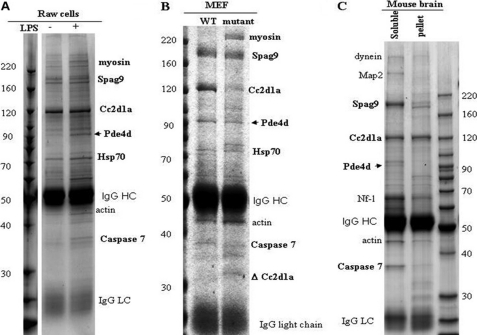
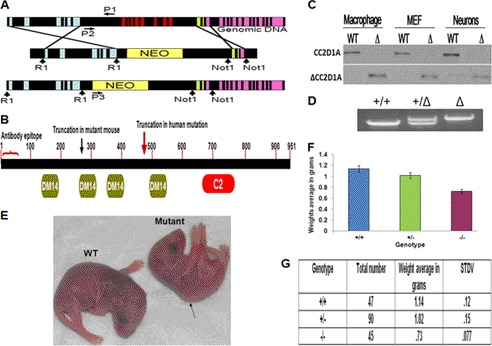
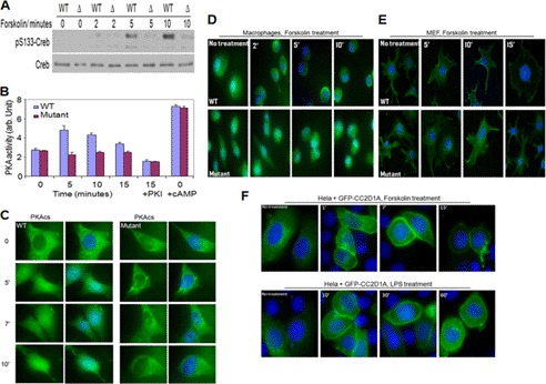
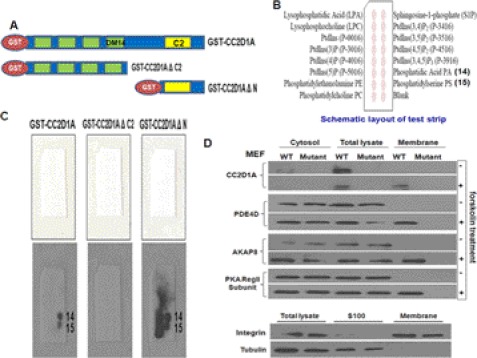
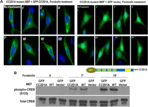
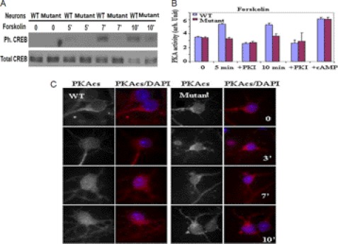
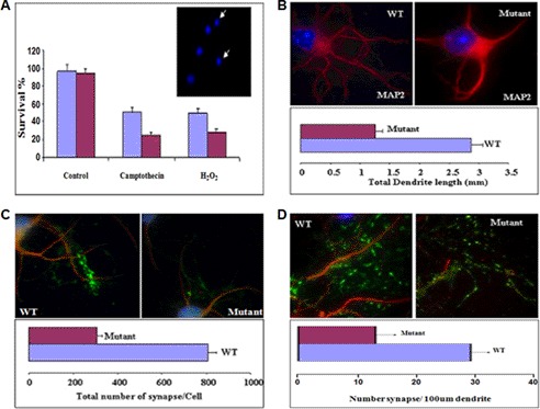
Mutation of the coiled-coil and C2 domain-containing 1A (CC2D1A) gene, which encodes a C2 domain and DM14 domain-containing protein, has been linked to severe autosomal recessive nonsyndromic mental retardation. Using a mouse model that produces a truncated form of CC2D1A that lacks the C2 domain and three of the four DM14 domains, we show that CC2D1A is important for neuronal differentiation and brain development. CC2D1A mutant neurons are hypersensitive to stress and have a reduced capacity to form dendrites and synapses in culture. At the biochemical level, CC2D1A transduces signals to the cyclic adenosine 3',5'-monophosphate (cAMP)-protein kinase A (PKA) pathway during neuronal cell differentiation. PKA activity is compromised, and the translocation of its catalytic subunit to the nucleus is also defective in CC2D1A mutant cells. Consistently, phosphorylation of the PKA target cAMP-responsive element-binding protein, at serine 133, is nearly abolished in CC2D1A mutant cells. The defects in cAMP/PKA signaling were observed in fibroblast, macrophage, and neuronal primary cells derived from the CC2D1A KO mice. CC2D1A associates with the cAMP-PKA complex following forskolin treatment and accumulates in vesicles or on the plasma membrane in wild-type cells, suggesting that CC2D1A may recruit the PKA complex to the membrane to facilitate signal transduction. Together, our data show that CC2D1A is an important regulator of the cAMP/PKA signaling pathway, which may be the underlying cause for impaired mental function in nonsyndromic mental retardation patients with CC2D1A mutation.


Figures

FIGURE 1.
FIGURE 1.
Proteomic analysis of Cc2d1a complexes identify cAMP signaling components as interacting proteins. Immunoprecipitations of Cc2d1a using an anti-Cc2d1a antibody from cell or tissue extracts under different conditions are shown. A, Raw cells that were cycling and treated with lipopolysaccharide (LPS, 1 μg/ml); B, MEF cells; C, mouse brain extracts. The immunocomplexes were resolved on a 4–20% SDS-polyacrylamide gel and stained with Coomassie Blue. The SDS-PAGE was divided into 12 regions; the bands were digested with trypsin, and the resulting peptides were analyzed with mass spectrometry to identify the proteins.
FIGURE 2.
FIGURE 2.
Cc2d1a protein structure and targeting strategy for CC2D1A knockouts and phenotypes of the ΔCC2D1A mutant mice. A, schematic of the endogenous CC2D1A gene and the result of homologous recombination with the targeting vector. Disruption resulted in the deletion of exons 7–14 by homologous recombination with the targeting vector. Colored vertical boxes represent exons. Vertical arrows indicate restriction enzyme sites. P1, P2, and P3 are the common, wild-type, and mutant primers, respectively, used for genotyping by PCR. B, schematic of the conserved domains of the Cc2d1a protein, the C2 and the DM14 domains, the truncated form of the Cc2d1a protein expressed in ΔCC2D1A mutant mice, and the truncated form of the Cc2d1a protein expressed in human NSMR patients. The antibody against Cc2d1a recognizes the first 50 amino acids at the N terminus, thereby labeling full-length and all truncation mutants. C, Western blotting shows full-length endogenous Cc2d1a protein (120 kDa) in the wild-type cells (top) and the truncated Cc2d1a protein (lower) expressed only in cells from the mutant mice (Δ)(∼36 kDa). Macrophages, MEF, and hippocampal neurons were cultured from littermates wild-type and mutant (Δ). Newborn (P0) mice were collected, washed with PBS, and lysed in 1× RIPA buffer with protease inhibitors. Fifteen μg of protein was loaded from each lysate. D, PCR genotyping demonstrating the ∼2-kb wild type (+/+), heterozygous (+/−), and ∼8-kb mutant (−/−, Δ) genotype fragments. E, picture of P0 wild-type and mutant mice showing the difference in size between wild-type and mutant. The picture also displays the mutant mouse hunchback phenotype. F, diagram shows the difference in the average weight between wild-type, heterozygous, and mutant mice. (n = 180, p < 0.0001). G, distributions of the genotype and the body weight of P0 mice. STDV refers to means ± S.D.
FIGURE 3.
FIGURE 3.
Cc2d1a is required for activation of protein kinase A and subcellular localization of the Cc2d1a protein in response to increases in cAMP. A, Western blot of Ser-133 phosphorylation of CREB in response to forskolin treatment in WT and mutant MEF cells. B, in vitro PKA activity stimulated with forskolin in MEF cells. C, indirect immunofluorescence demonstrating PKAcs translocation into the nucleus in response to forskolin treatment in WT and mutant MEF cells. D and E, immunostaining show the accumulation of Cc2d1a protein toward the cell periphery in both macrophages and MEF cells after stimulation with forskolin. This localization phenotype is not shown in CC2D1A mutant cells neither before nor after forskolin treatment. F, images of HeLa cells show the accumulation of the transfected GFP-Cc2d1a fusion protein toward the cell periphery after stimulation with LPS (1 μg/ml) or forskolin (50 μm). In all cases, cells were fixed and analyzed by immunofluorescence using an anti-Cc2d1a antibody and DAPI to stain the nuclei. Deacon microscopy was used for imaging.
FIGURE 4.
FIGURE 4.
Cc2d1a is required for re-localization of the PKA signaling module to the plasma membrane, and Cc2d1a C2 domain binds phospholipids. A, Western blot showing the presence of Cc2d1a, PDE4D, AKAP8, and PKA regulatory subunit II in wild-type MEF cell membrane fractions only after stimulation and not in CC2D1A mutant MEF cells either before or after stimulation. Crude membranes were purified from WT and CC2D1A mutant MEF cells after stimulation for 5 min. Total cell lysates, crude membrane, and cytosol fractions were analyzed by Western blots to estimate shifts in the localization of proteins. Tubulin and membrane marker proteins were used to show the purity of membrane fractions. B, Western blot showing GST-Cc2d1a and GST-Cc2d1aΔN but not GST-Cc2d1aΔC fusion proteins bind PA and PS. Phospholipid binding assay and Western blots are used to determine the phospholipids types that Cc2d1a C2 domain can bind. Nonspecific protein binding was blocked by incubating the PIP MicroStrip membranes in TBST with 3% fatty acid-free BSA. The membranes were then incubated in 0.5 μg/ml GST-Cc2d1a, of GST-Cc2d1a ΔN, or GST-Cc2d1a ΔC2 separately in TBST plus 3% fatty acid BSA overnight at 4 °C. The membrane strips were then incubated with anti-GST mouse monoclonal antibody and anti-mouse IgG horseradish peroxidase-linked antibody, respectively. In the end, membranes were developed using the ECL detection system. C, schematic diagrams of a PIP MicroStrip membrane showing the types of lipid spotted on the membrane. D, schematic diagram of GST-Cc2d1a, GST-Cc2d1aΔN, and GST-Cc2d1aΔC fusion proteins constructs.
FIGURE 5.
FIGURE 5.
Cc2d1a expression rescues the CREB phosphorylation phenotype in CC2D1A mutant MEF cells. A, Western blots show that GFP-Cc2d1a expression rescues the early CREB phosphorylation defect in CC2D1A mutant MEF cells to the wild-type level. GFP-Cc2d1a fusion protein was expressed in CC2D1A mutant MEF cells. The transfected mutant cells, along with the wild-type MEF cells, were stimulated with forskolin. GFP vector (control) expression did not rescue the early CREB phosphorylation defect in the CC2D1A mutant cells to the wild-type level, whereas GFP-Cc2d1a expression rescued the phenotype. B, images of CC2D1A mutant MEF cells show the accumulation of the expressed GFP-Cc2d1a fusion protein toward the cell periphery after stimulation with forskolin, whereas GFP protein alone (control) does not.
FIGURE 6.
FIGURE 6.
Cc2d1a regulates PKA activity in the neuronal system. A, Western blot of Ser-133 phosphorylation of CREB in response to forskolin treatment in WT and CC2D1A mutant neurons. B, in vitro PKA activity assay showing the increase in PKA activity only in WT and not in CC2D1A mutant neurons stimulated with forskolin. C, immunostaining demonstrating PKA catalytic subunit translocation into the nucleus in response to forskolin treatment in WT but not CC2D1A mutant neurons.
FIGURE 7.
FIGURE 7.
Cc2d1a is important for neuronal survival and plasticity. A, immunostaining and dendrite length measurements showing that total dendrite length per cell is lower in CC2D1A mutant neurons compared with WT. Hippocampal neurons were cultured for 14 days, fixed, and stained with antibodies to MAPII. Dendrites were visualized by immunofluorescence microscopy. Fluorescent images were obtained and analyzed to measure total dendrite length per cell (p < 0.001). B, survival assay and immunostaining showing that the survival rate under oxidative stress is compromised in CC2D1A mutant neurons. Hippocampal neurons were cultured for 36 h, and stress was introduced by camptothecin (10 μm) or H2O2 (30 μm) treatment. Then the neurons were fixed and stained with Hoechst or DAPI to score healthy, intact nuclei as an estimate of cell survival. (p < 0.002). Immunostaining and synapse number assay showed that the total synaptic number (C) and the synaptic density (D) is lower in the CC2D1A mutant neurons compared with WT neurons. Hippocampal neurons were cultured for 14 days, fixed, and stained with anti-MAPII and anti-V GlutII antibodies. The number of synapses was counted per cell. Number of synapses per length of dendrite (100 μm) was also calculated. (p < 0.002).

References

    1. Basel-Vanagaite L. (2007) Genetics of autosomal recessive nonsyndromic mental retardation. Recent advances. Clin. Genet. 72, 167–174 - PubMed
    1. Ropers H. H. (2006) X-linked mental retardation. Many genes for a complex disorder. Curr. Opin. Genet. Dev. 16, 260–269 - PubMed
    1. Raymond F. L., Tarpey P. (2006) The genetics of mental retardation. Hum. Mol. Genet. 15, R110–R116 - PubMed
    1. Basel-Vanagaite L., Attia R., Yahav M., Ferland R. J., Anteki L., Walsh C. A., Olender T., Straussberg R., Magal N., Taub E., Drasinover V., Alkelai A., Bercovich D., Rechavi G., Simon A. J., Shohat M. (2006) The CC2D1A, a member of a new gene family with C2 domains, is involved in autosomal recessive nonsyndromic mental retardation. J. Med. Genet. 43, 203–210 - PMC - PubMed
    1. Kikkawa U., Kishimoto A., Nishizuka Y. (1989) The protein kinase C family. Heterogeneity and its implications. Annu. Rev. Biochem. 58, 31–44 - PubMed

MeSH terms