

Glomus tumor of the stomach: a clinicopathologic analysis of 10 cases and review of the literature
- PMID: 22375171
- PMCID: PMC3286739
- DOI: 10.5009/gnl.2012.6.1.52
Glomus tumor of the stomach: a clinicopathologic analysis of 10 cases and review of the literature
Abstract
Background/aims: Gastric glomus tumors are extremely rare, and presurgical confirmation is often impossible. The identification of clinical and radiologic characteristics of this tumor type is important for preoperative diagnosis and treatment planning.
Methods: In this study, we analyzed 10 cases of gastric glomus tumors resected at a single institute over 9 years.
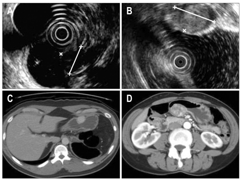
Results: Eight of the patients were men and 2 were women, with a mean age of 49 years. Five patients presented with abdominal discomfort or pain, 1 presented with anemia, and the remaining 4 cases were found incidentally during endoscopic examinations. The most common location of the tumor was the antrum (n=7), followed by the low (n=2) and high body (n=1). Although the endoscopic ultrasonography findings were variable, contrast-enhanced computed tomography generally showed a strong homogeneous enhancement. The resected tumors were well-demarcated solid masses with sizes ranging from 1.0 to 3.6 cm. Microscopically, the masses were composed of abundant vascular channels with clusters of uniform and round glomus cells. There was no evidence of recurrence after complete surgical resection.
Conclusions: Gastric glomus tumors are unusual, distinct lesions that should be considered in the differential diagnosis of a gastric submucosal mass. Unlike their deep soft tissue counterparts, most glomus tumors in the stomach are benign.
Keywords: Endoscopy; Glomus tumor; Pathology; Radiology; Stomach.
Conflict of interest statement
No potential conflict of interest relevant to this article was reported.
Figures


References
-
- Tsuneyoshi M, Enjoji M. Glomus tumor: a clinicopathologic and electron microscopic study. Cancer. 1982;50:1601–1607. - PubMed
-
- Miettinen M, Paal E, Lasota J, Sobin LH. Gastrointestinal glomus tumors: a clinicopathologic, immunohistochemical, and molecular genetic study of 32 cases. Am J Surg Pathol. 2002;26:301–311. - PubMed
-
- Kang JH, Lim JS, Kim JH, et al. Role of EUS and MDCT in the diagnosis of gastric submucosal tumors according to the revised pathologic concept of gastrointestinal stromal tumors. Eur Radiol. 2009;19:924–934. - PubMed
-
- Polkowski M. Endoscopic ultrasound and endoscopic ultrasound-guided fine-needle biopsy for the diagnosis of malignant submucosal tumors. Endoscopy. 2005;37:635–645. - PubMed
-
- Lee MJ, Lim JS, Kwon JE, et al. Gastric true leiomyoma: computed tomographic findings and pathological correlation. J Comput Assist Tomogr. 2007;31:204–208. - PubMed
LinkOut - more resources
Full Text Sources
