. 2012 May;53(5):918-928.
doi: 10.1194/jlr.M019075. Epub 2012 Feb 29.

Cell density-dependent reduction of dihydroceramide desaturase activity in neuroblastoma cells

Affiliations

Cell density-dependent reduction of dihydroceramide desaturase activity in neuroblastoma cells

Stefka D Spassieva et al. J Lipid Res. 2012 May.

Abstract

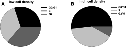
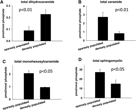
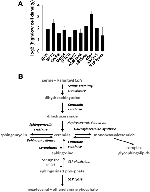
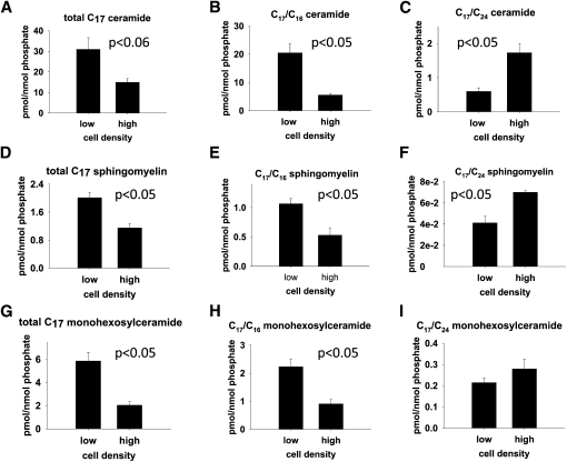
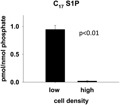
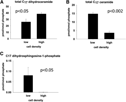
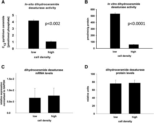
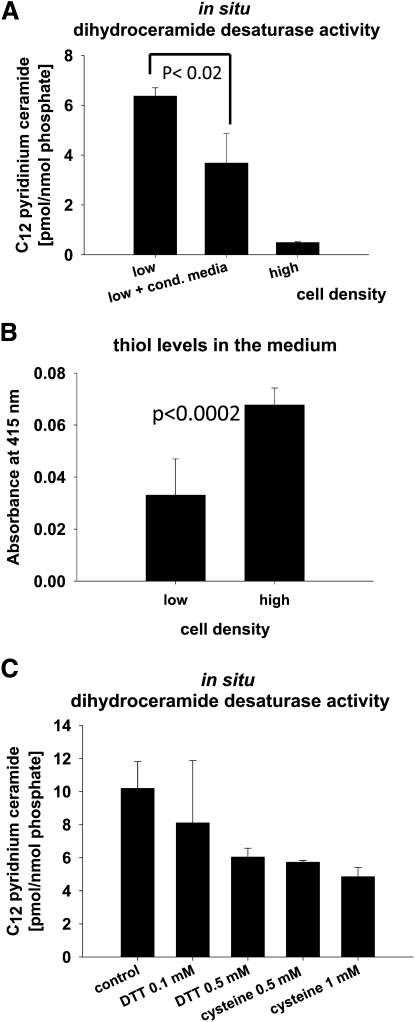
We applied a metabolic approach to investigate the role of sphingolipids in cell density-induced growth arrest in neuroblastoma cells. Our data revealed that sphingolipid metabolism in neuroblastoma cells significantly differs depending on the cells' population context. At high cell density, cells exhibited G0/G1 cell-cycle arrest and reduced ceramide, monohexosylceramide, and sphingomyelin, whereas dihydroceramide was significantly increased. In addition, our metabolic-labeling experiments showed that neuroblastoma cells at high cell density preferentially synthesized very long chain (VLC) sphingolipids and dramatically decreased synthesis of sphingosine-1-phosphate (S1P). Moreover, densely populated neuroblastoma cells showed increased message levels of both anabolic and catabolic enzymes of the sphingolipid pathway. Notably, our metabolic-labeling experiments indicated reduced dihydroceramide desaturase activity at confluence, which was confirmed by direct measurement of dihydroceramide desaturase activity in situ and in vitro. Importantly, we could reduce dihydroceramide desaturase activity in low-density cells by applying conditional media from high-density cells, as well as by adding reducing agents, such as DTT and L-cysteine to the media. In conclusion, our data suggest a role of the sphingolipid pathway, dihydroceramides desaturase in particular, in confluence-induced growth arrest in neuroblastoma cells.


Figures

Fig. 1.
Fig. 1.
High cell density results in growth arrest in neuroblastoma cells. SMS-KCNR cells grown at (A) low (50%) and (B) high (90%) cell densities were harvested, stained with PI, and subjected to cell-cycle analysis by flow cytometry. The charts represent the mean from two independent experiments.
Fig. 2.
Fig. 2.
MS analyses of cellular sphingolipid levels at low and high cell densities. SMS-KCNR cells were cultured at low (50%) and high (90%) cell densities. Cells were collected 48 h later; lipids were extracted and sphingolipid levels were determined by LC/MS analyses. Sphingolipid levels were normalized to cellular lipid phosphate. Error bars represent four independent experiments. (A) Dihydroceramide. (B) Ceramide. (C) Monohexosylceramide. (D) Sphingomyelin.
Fig. 3.
Fig. 3.
Upregulation of sphingolipid metabolic enzymes at high cell density. (A) RT-PCR results from RNAs isolated from cells grown until low (50%) and high (90%) cell densities. Error bars represent the results from three independent experiments (for SPT1, SPT2, SMS2, and aCer, n = 2). (B) Schematic representation of the sphingolipid pathway. Enzymes shown in part A to have increased message levels are in bold. alkCer1, alkaline ceramidase 1; aCer, acid ceramidase; CerS, ceramide synthase; aSMase, acid sphingomyelinase; nSMase2, neutral sphingomyelinase 2; SMS2, sphingomyelin synthase 2; SPT, serine palmitoyltransferase; UGCG, glucosylceramide synthase.
Fig. 4.
Fig. 4.
Metabolic labeling with C17 sphingosine of neuroblastoma cells cultured at low and high cell densities. SMS-KCNR cells were cultured at low (50%) and high (90%) cell densities. After 48 h, cells were labeled with 1 µM C17 sphingosine for 30 min. Lipids were extracted, and C17 sphingolipid species were measured by LC/MS analyses. C17 sphingolipid levels were normalized to cellular lipid phosphate. Error bars represent the results from three independent experiments. (A) Total C17 ceramide (the data used for calculating the total C17 ceramide is shown in supplementary Table I). (B) C17/C16 ceramide. (C) C17/C24 ceramide. (D) Total C17 sphingomyelin (the data used for calculating the total C17 sphingomyelin is shown in supplementary Table II). (E) C17/C16 sphingomyelin. (F) C17/C24 sphingomyelin. (G) Total C17 monohexosylceramide (the data used for calculating the total C17 monohexosylceramide is shown in supplementary Table III). (H) C17/C16 monohexosylceramide. (I) C17/C24 monohexosylceramide.
Fig. 5.
Fig. 5.
At high cell density, the neuroblastoma cells significantly decrease sphingosine-1 phosphate synthesis. Cells were cultured, labeled, and lipids were extracted and measured as in Fig. 4. C17 sphingosine-1-phosphate levels were normalized to cellular lipid phosphate. Error bars represent the results from three independent experiments.
Fig. 6.
Fig. 6.
Metabolic labeling with C17 dihydrosphingosine of neuroblastoma cells cultured at different cell densities (total). SMS-KCNR cells were cultured at low (50%) and high (90%) cell densities. After 48 h, cells were labeled with 1 µM C17 dihydrosphingosine for 30 min. Lipids were extracted, and C17 sphingolipid species were measured by LC/MS analyses. C17 sphingolipid levels were normalized to cellular lipid phosphate. Error bars represent the results from three independent experiments. (A) Total C17 dihydroceramide (the data used for calculating the total C17 dihydroceramide is shown in supplementary Table IV). (B) total C17 ceramide (the data used for calculating the total C17 ceramide is shown in supplementary Table V). (C) C17 dihydrosphingosine-1-phosphate.
Fig. 7.
Fig. 7.
Metabolic labeling with C17 dihydrosphingosine of neuroblastoma cells cultured at different cell densities (individual species). Cells were cultured and labeled, and lipids were extracted and measured as in Fig. 6. C17 sphingolipid levels were normalized to cellular lipid phosphate. Error bars represent the results from three independent experiments. (A) C17/C14 dihydroceramide. (B) C17/C16 dihydroceramide. (C) C17/C24 dihydroceramide. (D) C17/C26 dihydroceramide. (E) C17/C14 ceramide. (F) C17/C16 ceramide. (G) C17/C24 ceramide. (H) C17/C26 ceramide.
Fig. 8.
Fig. 8.
In neuroblastoma cells, dihydroceramide desaturase activity is reduced at high cell density. SMS-KCNR neuroblastoma cells were cultured at low (50%) and high (90%) cell densities for 48 h. (A) For the in situ dihydroceramide desaturase activity. An amount of 500 nM C12-dhCCPS, a dihydroceramide desaturase substrate, was added to the cells for 30 min. The product of the dihydroceramide desaturase reaction, C12-CCPS, was extracted from the collected cell pellets and quantified by MS. Results were normalized to the cellular lipid phosphate. Error bars represent the results from three independent experiments. (B) In vitro dihydroceramide desaturase activity. Error bars represent the results from four independent experiments. (C) RT-PCR results with dihydroceramide desaturase-specific primers on RNA isolated from low and high cell density cultured neuroblastoma cells. Expression of dihydroceramide desaturase was normalized to GAPDH. Error bars represent the results from three independent experiments. (D) Quantification of Western blot analyses on lysates from low and high cell density cultured neuroblastoma cells with dihydroceramide desaturase-specific antibody (Image J). Error bars represent the results from four independent experiments.
Fig. 9.
Fig. 9.
Reducing factors in the medium caused decrease in dihydroceramide desaturase activity. (A) SMS-KCNR neuroblastoma cells were cultured at low (50%) and high (90%) cell densities for 48 h. During the last 7 h of the experiment, in a subset of the samples cultured at low cell density, the growth media was replaced with conditioned media derived from cells cultured at high cell density. The in situ dihydroceramide desaturase activity was measured by adding 500 nM C12-dhCCPS (a dihydroceramide desaturase substrate) to the cells for 30 min. The product of the dihydroceramide desaturase reaction, C12-CCPS, was extracted from the collected cell pellets and quantified by MS. Results were normalized to the cellular lipid phosphate. Error bars represent the results from three independent experiments. (B) A standard “Ellman's test” was used for quantitation of the free thiols in the medium of SMS-KCNR cells cultures at low and high cell densities for 48 h. Error bars represent the standard deviation from six independent samples (two experiments). (C) SMS-KCNR cells were cultured at low cell density for 24 h. Reducing agents, DTT or l-cysteine, were added to the media and the cells were cultured for additional 24 h. The in situ dihydroceramide desaturase activity was measured as describe above (see part A). Error bars represent range of two independent experiments.

Similar articles

Cited by

References

    1. Hannun Y. A., Obeid L. M. 2008. Principles of bioactive lipid signalling: lessons from sphingolipids. Nat. Rev. Mol. Cell Biol. 9: 139–150 - PubMed
    1. Nelson P. J., Daniel T. O. 2002. Emerging targets: molecular mechanisms of cell contact-mediated growth control. Kidney Int. 61: S99–S105 - PubMed
    1. Hall P. A., Lane D. P. 1994. Genetics of growth arrest and cell death: key determinants of tissue homeostasis. Eur. J. Cancer. 30A: 2001–2012 - PubMed
    1. Kopitz J., Muhl C., Ehemann V., Lehmann C., Cantz M. 1997. Effects of cell surface ganglioside sialidase inhibition on growth control and differentiation of human neuroblastoma cells. Eur. J. Cell Biol. 73: 1–9 - PubMed
    1. Rösner H., Greis C., Rodemann H. P. 1990. Density-dependent expression of ganglioside GM3 by human skin fibroblasts in an all-or-none fashion, as a possible modulator of cell growth in vitro. Exp. Cell Res. 190: 161–169 - PubMed

Publication types