. 2012 Jun;53(6):1153-62.
doi: 10.1194/jlr.M024455. Epub 2012 Feb 29.

Cholesterol level influences opioid signaling in cell models and analgesia in mice and humans

Affiliations

Cholesterol level influences opioid signaling in cell models and analgesia in mice and humans

Hui Zheng et al. J Lipid Res. 2012 Jun.

Abstract

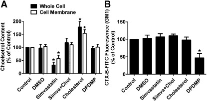
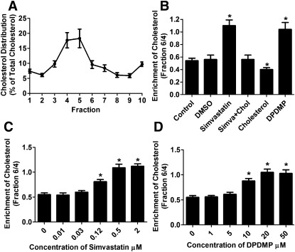
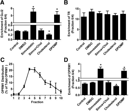
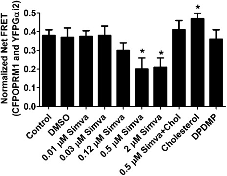
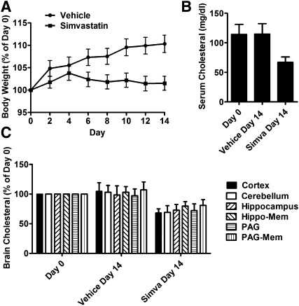
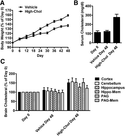
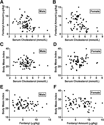
Cholesterol regulates the signaling of µ-opioid receptor in cell models, but it has not been demonstrated in mice or humans. Whether cholesterol regulates the signaling by mechanisms other than supporting the entirety of lipid raft microdomains is still unknown. By modulating cholesterol-enriched lipid raft microdomains and/or total cellular cholesterol contents in human embryonic kidney cells stably expressing µ-opioid receptor, we concluded that cholesterol stabilized opioid signaling both by supporting the lipid raft's entirety and by facilitating G protein coupling. Similar phenomena were observed in the primary rat hippocampal neurons. In addition, reducing the brain cholesterol level with simvastatin impaired the analgesic effect of opioids in mice, whereas the opioid analgesic effect was enhanced in mice fed a high-cholesterol diet. Furthermore, when the records of patients were analyzed, an inverse correlation between cholesterol levels and fentanyl doses used for anesthesia was identified, which suggested the mechanisms above could also be applicable to humans. Our results identified the interaction between opioids and cholesterol, which should be considered in clinics as a probable route for drug-drug interaction. Our studies also suggested that a low cholesterol level could lead to clinical issues, such as the observed impairment in opioid functions.


Figures

Fig. 1.
Fig. 1.
Simvastatin and DPDMP affect membrane composition. HEKOPRM1 cells were treated with PBS (Control), DMSO (DMSO), 0.5 μM simvastatin (Simvastatin), 0.5 μM simvastatin with 40 ng/ml cholesterol (Simva+Chol), 40 ng/ml cholesterol (Cholesterol), or 20 μM DPDMP (DPDMP) for 12 h. The cholesterol levels were determined in whole-cell lysis and on cell membrane in (A). The amounts of membrane GM1 were determined by FACS with CTX-B-FITC in (B). Data were analyzed by one-way ANOVA with Dunnett's posthoc test. Error bars and asterisks represent SD (n ≥ 4) and significant difference from Control, respectively.
Fig. 2.
Fig. 2.
Simvastatin and DPDMP affect cholesterol distribution on membrane. (A) Cell membrane of HEKOPRM1 cells were subjected for the continuous sucrose gradient as described in Materials and Methods. The cholesterol amounts were determined in each fraction and normalized against the whole cholesterol amount in the first 10 fractions. (B) Cells were treated as in Fig. 1A. The ratios of cholesterol amounts in fraction 6 to those in fraction 4 were summarized. (C, D) HEKOPRM1 cells were treated with indicated concentration of simvastatin (C) or DPDMP (D) for 12 h. The ratios of cholesterol amounts in fraction 6 to those in fraction 4 were summarized. Data were analyzed by one-way ANOVA with Dunnett's posthoc test. Error bars and asterisks represent SD (n ≥ 4) and significant difference from Control, respectively.
Fig. 3.
Fig. 3.
Simvastatin and DPDMP disrupt lipid raft microdomains. HEKOPRM1 cells were treated with PBS (Control), DMSO (DMSO), 0.5 μM simvastatin (Simvastatin), 0.5 μM simvastatin with 40 ng/ml cholesterol (Simva+Chol), 40 ng/ml cholesterol (Cholesterol), or 20 μM DPDMP (DPDMP) for 12 h. Cell membrane was subjected for the continuous sucrose gradient. The ratios of protein amounts in fraction 6 to those in fraction 4 were calculated. Lipid raft marker of Gαi2 was summarized in (A) and nonraft marker TR was summarized in (B). The distribution of OPRM1 on cell membrane was illustrated by the percentage amounts of OPRM1 in the first 10 fractions in the continuous sucrose gradient under “Control” condition (C). The ratios of OPRM1 amounts in fraction 6 to those in fraction 4 were summarized in (D). Data were analyzed by one-way ANOVA with Dunnett's posthoc test. Error bars and asterisks represent SD (n ≥ 4) and significant difference from Control, respectively.
Fig. 4.
Fig. 4.
Simvastatin and DPDMP affect Gαi2 coupling differentially. HEKOPRM1 cells were transfected with CFPOPRM1 and YFPGαi2. One day after the transfection, the cells were treated PBS (Control), DMSO (DMSO), 0.01 μM simvastatin (0.01 µM Simva), 0.03 μM simvastatin (0.03 µM Simva), 0.12 μM simvastatin (0.12 µM Simva), 0.5 μM simvastatin (0.5 µM Simva), 2 μM simvastatin (2 µM Simva), 0.5 μM simvastatin with 40 ng/ml cholesterol (Simva+Chol), 40 ng/ml cholesterol (Cholesterol), or 20 μM DPDMP (DPDMP) for 12 h. The normalized net FRET was determined on the cell membrane as described in Materials and Methods. Data were analyzed by one-way ANOVA with Dunnett's posthoc test. Error bars and asterisks represent SD (n ≥ 12) and significant difference from Control, respectively.
Fig. 5.
Fig. 5.
Simvastatin modulates opioid analgesia. Two groups were injected with 10 mg/kg simvastatin or vehicle twice per day for 14 days. (A) Body weights of each mouse were measured every two days and normalized to those in day 0. (B) Cholesterol levels were determined on day 0 and day 14 in serum. (C) Cholesterol levels were determined on day 0 and day 14 in cortex, cerebellum, hippocampus, the cell membrane of neurons in hippocampal (Hippo-Mem), PAG, and the cell membrane of neurons in PAG (PAG-Mem). Data were analyzed by one-way ANOVA with Dunnett's posthoc test. Error bars represent SD (n ≥ 10) and significant difference from day 0, respectively.
Fig. 6.
Fig. 6.
High-cholesterol diet modulates opioid analgesia. Two groups were fed with control diet and high-cholesterol diet for 48 days. (A) Body weights of each mouse were measured every six days and normalized to those in day 0. (B) Cholesterol levels were determined on day 0 and day 48 in serum. (C) Cholesterol levels were determined on day 0 and day 48 in different regions in brain as in Fig. 3C. Data were analyzed by one-way ANOVA with Dunnett's posthoc test. Error bars represent SD (n ≥ 10) and significant difference from day 0, respectively.
Fig. 7.
Fig. 7.
Serum cholesterol levels inversely correlate with the amounts of fentanyl used for anesthesia. The total serum cholesterol levels were plotted against the fentanyl amounts used before the surgery, Male in (A) and Female in (B). The BMIs were plotted against the total serum cholesterol levels, Male in (C) and Female in (D). The BMIs were plotted against the fentanyl amounts used before the surgery, Male in (E) and Female in (F). The correlation was tested with two-tailed Pearson rank test. The Pearson's rank correlation coefficients and P values are listed in the text.

Similar articles

Cited by

References

    1. Chini B., Parenti M. 2004. G-protein coupled receptors in lipid rafts and caveolae: how, when and why do they go there? J. Mol. Endocrinol. 32: 325–338 - PubMed
    1. Helms J. B., Zurzolo C. 2004. Lipids as targeting signals: lipid rafts and intracellular trafficking. Traffic. 5: 247–254 - PubMed
    1. Ostrom R. S., Gregorian C., Drenan R. M., Xiang Y., Regan J. W., Insel P. A. 2001. Receptor number and caveolar co-localization determine receptor coupling efficiency to adenylyl cyclase. J. Biol. Chem. 276: 42063–42069 - PubMed
    1. Navratil A. M., Bliss S. P., Berghorn K. A., Haughian J. M., Farmerie T. A., Graham J. K., Clay C. M., Roberson M. S. 2003. Constitutive localization of the gonadotropin-releasing hormone (GnRH) receptor to low density membrane microdomains is necessary for GnRH signaling to ERK. J. Biol. Chem. 278: 31593–31602 - PubMed
    1. Li S., Couet J., Lisanti M. P. 1996. Src tyrosine kinases, Galpha subunits, and H-Ras share a common membrane-anchored scaffolding protein, caveolin. Caveolin binding negatively regulates the auto-activation of Src tyrosine kinases. J. Biol. Chem. 271: 29182–29190 - PMC - PubMed

Publication types

MeSH terms