. 2012 Jun;93(Pt 6):1316-1327.
doi: 10.1099/vir.0.040790-0. Epub 2012 Feb 29.

Virion endocytosis is a major target for murid herpesvirus-4 neutralization

Affiliations

Virion endocytosis is a major target for murid herpesvirus-4 neutralization

Daniel L Glauser et al. J Gen Virol. 2012 Jun.

Abstract

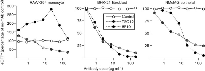
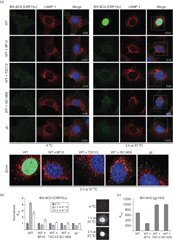
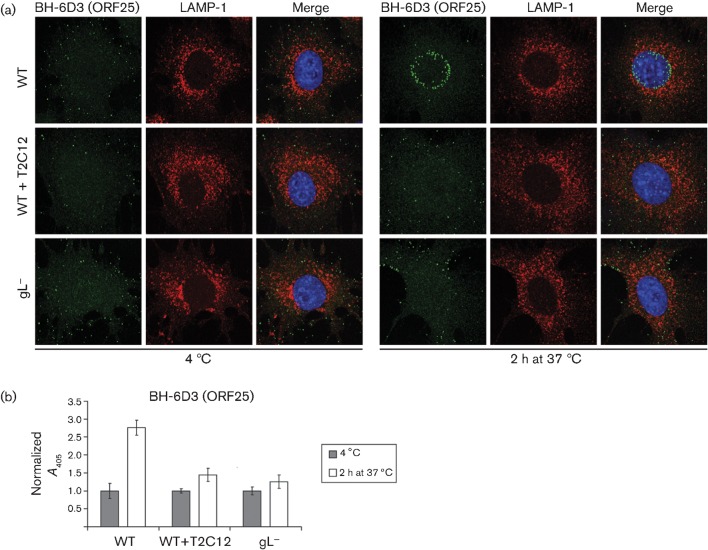
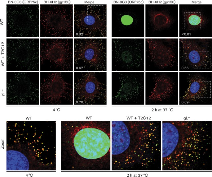
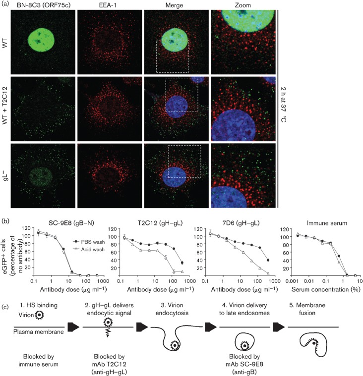
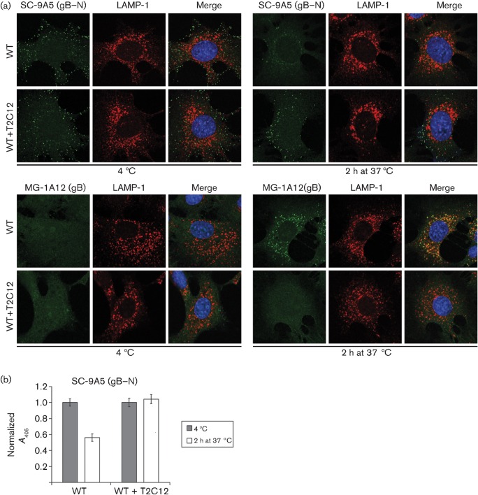
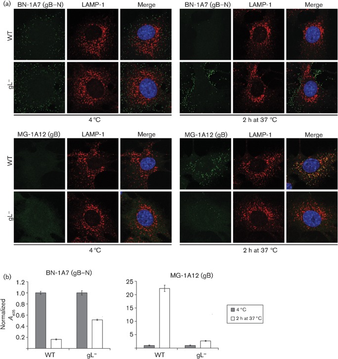
Herpesviruses consistently transmit from immunocompetent carriers, implying that their neutralization is hard to achieve. Murid herpesvirus-4 (MuHV-4) exploits host IgG Fc receptors to bypass blocks to cell binding, and pH-dependent protein conformation changes to unveil its fusion machinery only after endocytosis. Nevertheless, neutralization remains possible by targeting the virion glycoprotein H (gH)-gL heterodimer, and the neutralizing antibody responses of MuHV-4 carriers are improved by boosting with recombinant gH-gL. We analysed here how gH-gL-directed neutralization works. The MuHV-4 gH-gL binds to heparan sulfate. However, most gH-gL-specific neutralizing antibodies did not block this interaction; neither did they act directly on fusion. Instead, they blocked virion endocytosis and transport to the late endosomes, where membrane fusion normally occurs. The poor endocytosis of gH-gL-neutralized virions was recapitulated precisely by virions genetically lacking gL. Therefore, driving virion uptake appears to be an important function of gH-gL that provides a major target for antibody-mediated neutralization.


Figures

Fig. 1.
Fig. 1.
Comparison of virion neutralization by gH–gL-specific mAbs T2C12 and 8F10. MuHV-4 virions expressing eGFP from an intergenic EF1α promoter were incubated (2 h, 37 °C) with antibody dilutions, then divided between RAW-264 monocytes (3 p.f.u. per cell), BHK-21 fibroblasts (0.5 p.f.u. per cell) and NMuMG epithelial cells (0.5 p.f.u. per cell). After overnight incubation (37 °C) in the presence of 100 µg phosphonoacetic acid ml−1 to prevent secondary spread, the cells were analysed for eGFP expression by flow cytometry. The MHC class I-specific IgG2a mAb 28.14.8 provided a control.
Fig. 2.
Fig. 2.
Transport of neutralized virions. (a) Wild-type (WT) MuHV-4 virions (3 p.f.u. per cell) were left untreated or pre-incubated (2 h, 37 °C) with mAbs 8F10 (anti-gH–gL, IgG2a), T2C12 (anti-gH–gL, IgG2a) or SC-9E8 (anti-gB, IgG2a) (400 µg ml−1) before binding to NMuMG cells (2 h, 4 °C). For comparison, other NMuMG cells were similarly exposed to gL virions (50 p.f.u. per cell, so as to get equivalent binding). The cells were then washed with PBS and either fixed immediately or first further incubated (2 h, 37 °C) to allow virion endocytosis. The cells were then stained for the ORF75c virion tegument protein with mAb BN-8C3 (IgG1, green) and for the late endosomal marker LAMP-1 (red), and with DAPI (blue). Red/green co-localization appears as yellow. Equivalent data were obtained in a repeat experiment. In this and all subsequent figures, the data shown are fully representative of at least 100 cells examined. The confocal settings were the same for the corresponding images at 4 °C and after 2 h at 37 °C. The numbers give the fraction of green signal co-localizing with red signal. The zoomed images show in more detail the relationship between virions (green) and endosomes (red). (b) As in (a), cells were exposed to virions for 2 h at 4 °C, washed in PBS, then either analysed immediately or first incubated for 1 or 2 h at 37 °C, but antibody binding was detected with an IgG1-specific alkaline phosphatase-conjugated secondary antibody and incubation with p-nitrophenylphosphate substrate, and quantified by measuring A405. For each condition, the A405 was normalized to the value obtained at 4 °C. The bars show mean±sem values from six wells. The ORF75c signal after incubation at 37 °C was significantly higher for non-neutralized WT virions than for gL or neutralized WT virions (P<0.008 by Student’s t-test). The images show the distribution of ORF75c after virion binding at 4 °C, and after incubation at 37 °C for 1 or 2 h. (c) In a similar experiment to (b), virions were bound to cells for 2 h at 4 °C and detected with the gp150-specific IgG2b mAb BH-6H2 plus an alkaline phosphatase-conjugated IgG2b-specific secondary antibody. The bars show mean±sem A405 values from six wells. The signal with 8F10-neutralized virions was reduced significantly relative to other treatments (P<10−5). Equivalent data were obtained in a repeat experiment.
Fig. 3.
Fig. 3.
Capsid migration of T2C12-neutralized and gL virions. (a) WT MuHV-4 virions (3 p.f.u. per cell) were left untreated or pre-incubated (2 h, 37 °C) with mAb T2C12 (anti-gH–gL, IgG2a, 400 µg ml−1). WT (3 p.f.u. per cell) and gL (50 p.f.u. per cell to give equivalent binding) virions were then bound to NMuMG cells for 2 h at 4 °C. The cells were washed with PBS and either fixed immediately or first incubated (2 h, 37 °C) to allow virion endocytosis. All cells were then stained for the ORF25 virion capsid component with mAb BH-6D3 (IgG1, green), for the late endosomal marker LAMP-1 (red), and with DAPI (blue). Equivalent data were obtained in a repeat experiment. (b) Cells were exposed to WT virions with or without T2C12 neutralization or to gL virions as in (a), but antibody binding was detected with an alkaline phosphatase-conjugated IgG1-specific secondary antibody, p-nitrophenylphosphate substrate and A405. For each condition, the A405 was normalized to the value obtained at 4 °C. The bars show mean±sem values from six wells. After incubation at 37 °C, the non-neutralized WT signal was significantly higher than that of gL or T2C12-treated WT virions (P<0.0005 by Student’s t-test). Equivalent data were obtained in two further experiments.
Fig. 4.
Fig. 4.
Monitoring virion entry by envelope/tegument co-localization. WT MuHV-4 virions (3 p.f.u. per cell) were left untreated or pre-incubated (2 h, 37 °C) with mAb T2C12 (anti-gH–gL, IgG2a, 400 µg ml−1). WT (3 p.f.u. per cell) and gL (50 p.f.u. per cell to give equivalent binding) virions were then bound to NMuMG cells for 2 h at 4 °C. The cells were washed with PBS and either fixed immediately or first incubated (2 h, 37 °C) to allow virion endocytosis. They were then stained for the ORF75c tegument component with mAb BN-8C3 (IgG1, green), for the gp150 envelope protein with mAb BH-6H2 (IgG2b, red) and with DAPI (blue). The numbers give the fraction of green signal co-localizing with red signal. The zoomed images show this relationship in more detail.
Fig. 5.
Fig. 5.
gH–gL-neutralized virions show defective endocytosis. (a) WT MuHV-4 virions (3 p.f.u. per cell) were left untreated or incubated (2 h, 37 °C) with mAb T2C12 (anti-gH–gL, IgG2a, 400 µg ml−1). These and gL virions (50 p.f.u. per cell to give equivalent binding) were then bound to NMuMG cells (2 h, 4 °C). The cells were washed with PBS and incubated (2 h, 37 °C) to allow virion endocytosis, washed, fixed, and stained for the ORF75c virion tegument protein with mAb BN-8C3 (IgG1, green), for the early endosomal marker EEA-1 (red) and with DAPI (blue). Significant co-localization (yellow) was not observed. The zoomed images show virion and endosome distributions in more detail. (b) eGFP-expressing MuHV-4 virions (0.2 p.f.u. per cell) were left untreated or incubated (2 h, 37 °C) with mAb SE-9E8 (anti-gB, blocks fusion), mAb T2C12 (anti-gH–gL), mAb 7D6 (anti-gH–gL) or MuHV-4 immune serum (blocks binding). The virus was then added to BHK-21 cells and incubated (2 h, 37 °C) to allow virus endocytosis. The cells were then washed with PBS to remove unbound virions or with pH 3 citrate/phosphate buffer to inactivate non-endocytosed virions, then incubated (16 h, 37 °C) in complete medium with 100 µg phosphonoacetic acid ml−1 and analysed for viral eGFP expression by flow cytometry. Each point shows the mean±sem of two experiments. Comparison across each dilution by Fisher’s exact test established that acid washing reduced infection significantly for mAbs T2C12 and 7D6 (P<0.02), but not for mAb SC-9E8 or for immune serum (P>0.5). (c) A schematic diagram shows where neutralization seems to act during MuHV-4 entry. Immune sera block virion binding to the plasma membrane; mAb T2C12 blocks the endocytosis of bound virions, and so presumably prevents the delivery of an endocytic signal by its target, gH–gL; gB-directed neutralization blocks membrane fusion and so strands virions in late endosomes.
Fig. 6.
Fig. 6.
gH–gL-directed neutralization prevents conformation changes in gB. (a) WT MuHV-4 virions (3 p.f.u. per cell) were left untreated or pre-incubated (2 h, 37 °C) with mAb T2C12 (IgG2a, 400 µg ml−1), then bound to NMuMG cells (2 h, 4 °C). The cells were washed with PBS and either fixed immediately or first further incubated (2 h, 37 °C) to allow virion endocytosis, then stained with mAb SC-9A5 (IgG3), which is specific for pre-fusion gB, and with mAb MG-1A12 (IgG2a), which is specific for post-fusion gB (Glauser et al., 2011). SC-9A5 was detected with an Alexa Fluor 488-conjugated goat anti-mouse IgG3 pAb; MG-1A12 was conjugated directly to Alexa Fluor 488 (both green). The cells were also stained for LAMP-1 (red) and with DAPI (blue). (b) NMuMG cells were exposed to untreated or T2C12-neutralized WT virions as in (a), then stained for pre-fusion gB with mAb SC-9A5. Antibody binding was detected with alkaline phosphatase-conjugated IgG3-specific secondary antibody, p-nitrophenylphosphate substrate and A405. For each condition, the A405 was normalized to the value obtained at 4 °C. The bars show mean±sem values from six wells. The non-neutralized WT signal was reduced significantly after incubation at 37 °C (P<10−4 by Student’s t-test), whereas the neutralized WT signal was not changed significantly (P>0.58). Equivalent data were obtained in a repeat experiment.
Fig. 7.
Fig. 7.
gL virions also show little conformation change in gB. (a) NMuMG cells were incubated (2 h, 4 °C) with WT (3 p.f.u. per cell) or gL (50 p.f.u. per cell for equivalent binding) MuHV-4, then washed with PBS and either fixed immediately or first further incubated (2 h, 37 °C) to allow virion endocytosis. The cells were stained for pre-fusion gB with mAb BN-1A7 (IgG2a) or for post-fusion gB with mAb MG-1A12 (IgG2a) (both green), for LAMP-1 (red) and with DAPI (blue). Co-localization appears as yellow. Equivalent data were obtained in three further experiments. (b) Cells and viruses were incubated as in (a), then antibody binding was detected with an alkaline phosphatase-conjugated IgG2a-specific secondary antibody, p-nitrophenylphosphate substrate and A405. For each condition, the A405 was normalized to the value obtained at 4 °C. The bars show mean±sem values from six wells. After incubation at 37 °C, the WT BN-1A7 signal was reduced significantly relative to gL (P<10−9 by Student’s t-test) and the WT MG-1A12 signal was increased significantly (P<10−5). Equivalent data were obtained in a repeat experiment.

Similar articles

Cited by

References

    1. Barton E., Mandal P., Speck S. H. (2011). Pathogenesis and host control of gammaherpesviruses: lessons from the mouse. Annu Rev Immunol 29, 351–397 10.1146/annurev-immunol-072710-081639 - DOI - PubMed
    1. Chandran B. (2010). Early events in Kaposi’s sarcoma-associated herpesvirus infection of target cells. J Virol 84, 2188–2199 10.1128/JVI.01334-09 - DOI - PMC - PubMed
    1. Chesnokova L. S., Hutt-Fletcher L. M. (2011). Fusion of Epstein–Barr virus with epithelial cells can be triggered by αvβ5 in addition to αvβ6 and αvβ8, and integrin binding triggers a conformational change in glycoproteins gHgL. J Virol 85, 13214–13223 10.1128/JVI.05580-11 - DOI - PMC - PubMed
    1. Chesnokova L. S., Nishimura S. L., Hutt-Fletcher L. M. (2009). Fusion of epithelial cells by Epstein–Barr virus proteins is triggered by binding of viral glycoproteins gHgL to integrins αvβ6 or αvβ8. Proc Natl Acad Sci U S A 106, 20464–20469 10.1073/pnas.0907508106 - DOI - PMC - PubMed
    1. Chowdary T. K., Cairns T. M., Atanasiu D., Cohen G. H., Eisenberg R. J., Heldwein E. E. (2010). Crystal structure of the conserved herpesvirus fusion regulator complex gH–gL. Nat Struct Mol Biol 17, 882–888 10.1038/nsmb.1837 - DOI - PMC - PubMed

Publication types

MeSH terms

Substances

LinkOut - more resources