Comparative Study
. 1990 Nov;108(5):880-9.

Rejection of multivisceral allografts in rats: a sequential analysis with comparison to isolated orthotopic small-bowel and liver grafts

Affiliations
Comparative Study

Rejection of multivisceral allografts in rats: a sequential analysis with comparison to isolated orthotopic small-bowel and liver grafts

N Murase et al. Surgery. 1990 Nov.

Abstract

Multivisceral isografts and allografts were transplanted to Lewis rats, and the histopathologic changes were studied in the liver, intestine, and other constituent organs. Rats receiving isografts had indefinite survival with maintenance of weight. With multivisceral allografts (from Brown-Norway donors), the intestinal component was rejected more severely than the companion liver and with about the same severity as when intestinal transplantation was performed alone. Intestinal rejection in either circumstance was a lethal event, causing death in 10 to 12 days. The earliest (by day 4) and most intense cellular rejection was in the Peyer's patches and mesenteric lymph nodes. This was associated with or followed by cryptitis, epithelial cell necrosis, focal abscess formation, mural necrosis, and eventual perforation. Liver allografts transplanted alone or as part of multivisceral grafts also had histopathologic evidence of rejection, but this was self-limiting and spontaneously reversible when the liver was transplanted alone. Thus the Achille's heel of multivisceral grafts is the intestinal component that is not protected by the presence of the liver in the organ complex. Better immunosuppression should permit successful experimental and clinical transplantation of such grafts.


Figures
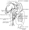
Fig. 1
Fig. 1
Technique of multivisceral transplantation. IVC, Inferior vena cava; SMA, superior mesenteric artery.
Fig. 2
Fig. 2
Body weight (mean ± SD) after transplantation for each of the operative procedures.
Fig. 3
Fig. 3
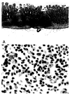
Small intestine from a multivisceral isograft. A, Peyer's patch on day 5; note intact villi and normal-appearing lymphoid tissue. (Hematoxylin and eosin stain; original magnification ×100.) B, High-power view of A shows small inactive lymphoid cells in the interfollicular T-cell zone (see arrow in A). The isografts remained normal thereafter. (Hematoxylin and eosin stain; original magnification ×1000.)
Fig. 4
Fig. 4
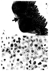
Small intestine from a small-bowel allograft. A, Peyer's patch on day 4 with mild expansion of the T-cell zone (arrow). (Hematoxylin and eosin stain; original magnification ×40.) B, High-power view of T- cell zone shown in A. Note the blastic transformation and compare with isograft in Fig. 3, B.
Fig. 5
Fig. 5
Small intestine from a small-bowel allograft. A, Peyer's patch region on day 7. Note the marked expantion of T-cell zones (arrowheads). (Hematoxylin and eosin stain; original magnification ×40.) B, High-power view of T-cell zone in A demonstrates immunoblastic and histocytic cells. (Hematoxylin and eosin stain; original magnification ×400.) C, The lymphatics in the lamina propria also became dilated on day 7 in allografts. (Hematoxylin and eosin stain; original magnification ×100).
Fig. 6
Fig. 6
Small intestine on day 9 from a small-bowel allograft. A, By day 9 the Peyer's patches were largely replaced with histiocytes. Focal and mucosal breakdown of the epithelium overlying the Peyer's patches was evident. The cellular population shown in Fig. 5, B is typical for this time. (Hematoxylin and eosin stain; original magnification ×100.) B, Cryptitis is seen in the allograft small-intestine epithelium, away from the Peyer's patches (arrows). (Hematoxylin and eosin stain; original magnification ×400).
Fig. 7
Fig. 7
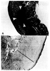
Intestine from a small-bowel allograft. A, By day 9, mucosal breakdown with leakage of intestinal contents and intramural abscess formation were evident (arrows). (Hematoxylin and eosin stain; original magnification ×40.) B, This was followed 2 to 3 days later by transmural necrosis (bar). (Hematoxylin and eosin stain; original magnification ×100.)

References

    1. Starzl TE, Kaupp HA, Brock DR, Butz GW, Linman JW. Homotransplantation of multiple visceral organs. Am J Surg. 1962;103:219–29. - PMC - PubMed
    1. Starzl TE, Rowe MI, Todo S, et al. Transplantation of multiple abdonimal viscera. JAMA. 1989;261:1449–57. - PMC - PubMed
    1. Williams JS, Sankary HN, Foster PF, Lowe J, Goldman GM. Splanchnic transplantation: an approach to the infant dependent on parentral nutrition who develops irreversible liver disease. JAMA. 1989;261:1458–62. - PubMed
    1. Kamada N. Experimental liver transplantation. 7-12. Boca Raton, FL: CRC Press; 1988. pp. 55–65.
    1. Jaffee R, Trager JDK, Zeevi A, et al. Multivisceral intestinal transplantation: surgical pathology. Pediatr Pathol. 1989;9:633–54. - PMC - PubMed

Publication types

MeSH terms

LinkOut - more resources