Comparative Study
. 2012 Apr;122(4):1553-66.
doi: 10.1172/JCI61143. Epub 2012 Mar 1.

c-Src and c-Abl kinases control hierarchic phosphorylation and function of the CagA effector protein in Western and East Asian Helicobacter pylori strains

Affiliations
Comparative Study

c-Src and c-Abl kinases control hierarchic phosphorylation and function of the CagA effector protein in Western and East Asian Helicobacter pylori strains

Doreen Mueller et al. J Clin Invest. 2012 Apr.

Abstract

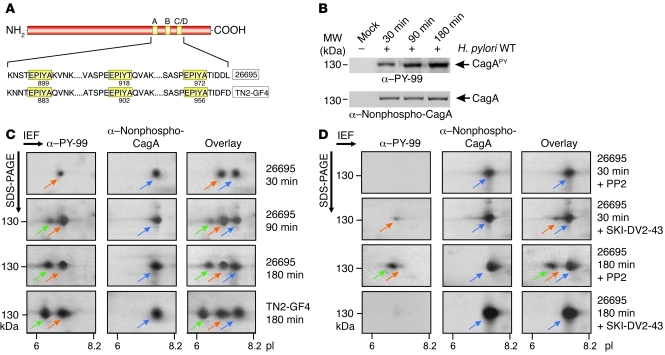
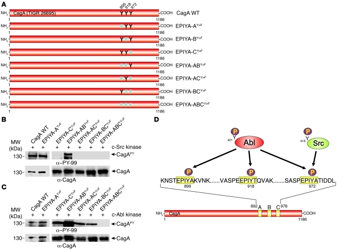
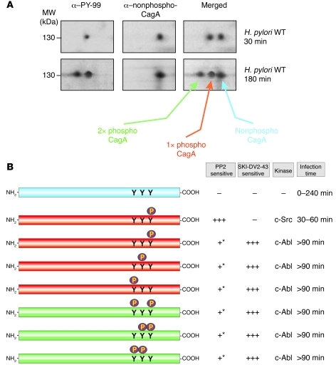
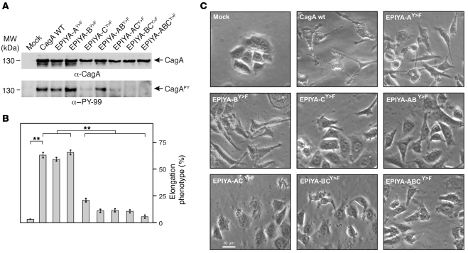
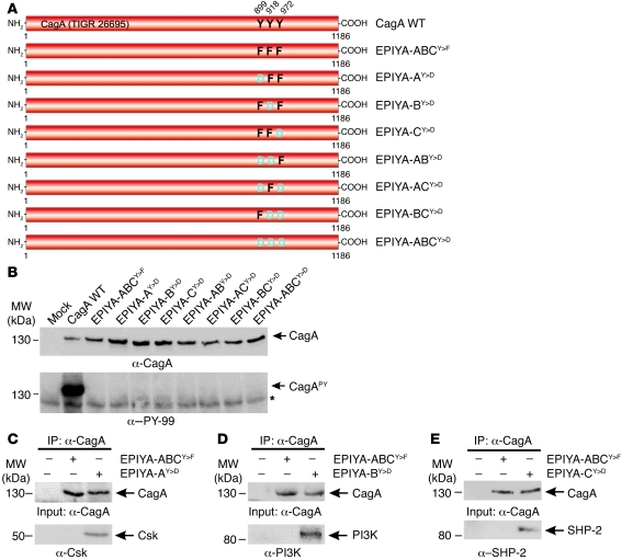
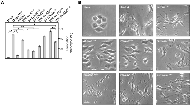
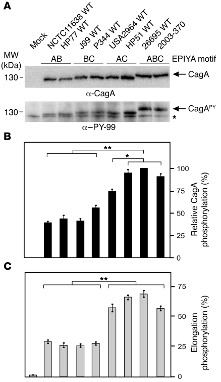
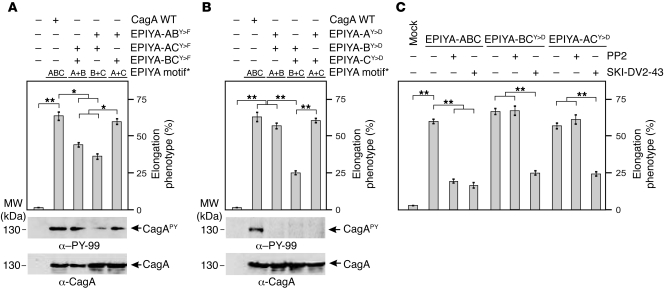
Many bacterial pathogens inject into host cells effector proteins that are substrates for host tyrosine kinases such as Src and Abl family kinases. Phosphorylated effectors eventually subvert host cell signaling, aiding disease development. In the case of the gastric pathogen Helicobacter pylori, which is a major risk factor for the development of gastric cancer, the only known effector protein injected into host cells is the oncoprotein CagA. Here, we followed the hierarchic tyrosine phosphorylation of H. pylori CagA as a model system to study early effector phosphorylation processes. Translocated CagA is phosphorylated on Glu-Pro-Ile-Tyr-Ala (EPIYA) motifs EPIYA-A, EPIYA-B, and EPIYA-C in Western strains of H. pylori and EPIYA-A, EPIYA-B, and EPIYA-D in East Asian strains. We found that c-Src only phosphorylated EPIYA-C and EPIYA-D, whereas c-Abl phosphorylated EPIYA-A, EPIYA-B, EPIYA-C, and EPIYA-D. Further analysis revealed that CagA molecules were phosphorylated on 1 or 2 EPIYA motifs, but never simultaneously on 3 motifs. Furthermore, none of the phosphorylated EPIYA motifs alone was sufficient for inducing AGS cell scattering and elongation. The preferred combination of phosphorylated EPIYA motifs in Western strains was EPIYA-A and EPIYA-C, either across 2 CagA molecules or simultaneously on 1. Our study thus identifies a tightly regulated hierarchic phosphorylation model for CagA starting at EPIYA-C/D, followed by phosphorylation of EPIYA-A or EPIYA-B. These results provide insight for clinical H. pylori typing and clarify the role of phosphorylated bacterial effector proteins in pathogenesis.


Figures

Figure 1
Figure 1. Analysis of CagAPY protein species during infection with H. pylori by 1-DE and 2-DE.
(A) CagA proteins vary in the C-terminal EPIYA phosphorylation sites depending on geographical origin. Western CagA proteins (e.g., strain 26695) contain the EPIYA-A, EPIYA-B, and EPIYA-C segments, while East Asian strains contain the EPIYA-A, EPIYA-B, and EPIYA-D segments (e.g., strain TN2-GF4). These motifs are tyrosine phosphorylation sites that can be phosphorylated by Abl and Src tyrosine kinases (–13). (B) AGS cells were infected for the indicated times with strain 26695. The resulting protein lysates were separated by 1-DE, and phosphorylation of injected CagA was examined using α–PY-99 and α-CagA antibodies (arrows). (C) Separation of CagA protein species from B by 2-DE. Depending on the time of infection, full-length CagAPY appeared as 1 spot (spot 1, red arrows, pI = 7.0) or 2 spots (spots 1 and 2; spot 2, green arrows, pI = 6.5) as indicated. The α-CagA antibody probe revealed a third spot (spot 3, blue arrows, pI = 7.5). Overlay of both exposures yielded 2 or 3 spots as shown. Strain TN2-GF4 exhibited the same pattern as 26695 (bottom). (D) Inhibition of Src with PP2 (10 μM) or Abl with SKI-DV2-43 (1 μM) revealed significant changes in spot intensity depending on the time of infection.
Figure 2
Figure 2. In vitro phosphorylation of CagA mutants by c-Abl or c-Src kinases.
(A) Site-directed Y>F mutagenesis of CagA EPIYA-A, EPIYA-B, and EPIYA-C in strain 26695. Single, double, and triple mutants were named as indicated. (B and C) Lysates of H. pylori expressing the mutated CagA EPIYA motifs were subjected to in vitro phosphorylation assays using recombinant c-Src kinase (B) or c-Abl kinase (C). Immunoblotting using α–PY-99 and α-CagA antibodies (arrows) indicated that both c-Src and c-Abl phosphorylated CagA in a different fashion. (D) Schematic diagram of the data, showing that c-Src only phosphorylated Y-972 in EPIYA-C, while Abl can phosphorylate Y-899, Y-918, and Y-972 in EPIYA-A, EPIYA-B, and EPIYA-C, respectively.
Figure 3
Figure 3. Modeling of phosphorylated CagA protein species detected in Figures 1 and 2.
(A) Representative 2-DE pictures of CagA protein species produced after 30 and 180 minutes of H. pylori infection. (B) Different possible phosphorylated CagA protein species induced during infection and their time-dependent sensitivity to pharmacological inhibition of Src and Abl kinases. Colors indicate the confirmed or proposed phosphorylation status of CagA motifs in each 2-DE spot. +++, strong effect; + moderate effect; –, no effect. Asterisks indicate that the slight inhibitory effect of PP2 on CagA protein species phosphorylated by c-Abl (>90 minutes) may be due to c-Src being an upstream component of the signal cascade leading to full c-Abl activation during infection.
Figure 4
Figure 4. Role of EPIYA motifs in CagA phosphorylation and AGS cell elongation during H. pylori infection.
(A) AGS cells were infected for 4 hours with CagA-expressing H. pylori strains as indicated. Phosphorylation of CagA was examined using α–PY-99 and α-CagA antibodies (arrows). (B) The number of elongated cells in each experiment was quantitated in triplicate in 10 different 0.25-mm2 fields. (C) Phase-contrast micrographs of AGS cells infected with the different strains as indicated. **P ≤ 0.001.
Figure 5
Figure 5. Generation of phosphomimetic CagA mutants and their interaction with host signaling factors during H. pylori infection.
(A) Site-directed Y>D mutagenesis of CagA EPIYA-A, EPIYA-B, and EPIYA-C to generate phosphomimetic mutants. Nontargeted tyrosines in adjacent EPIYA motifs were replaced by phenylalanines to avoid additional phosphorylation events per molecule. The resulting single, double, and triple mutants were named as indicated. (B) AGS cells were infected for 4 hours with CagA-expressing H. pylori strains as indicated. CagA phosphorylation was examined using α–PY-99 and α-CagA antibodies (arrows). All strains expressed similar amounts of CagA, but only H. pylori expressing WT CagA revealed a phosphorylation signal. The asterisk in the lower panel indicates antibody cross-reactivity with an unknown phosphorylated host cell protein. (CE) AGS cells were infected for 4 hours with CagA-expressing H. pylori strains as indicated, and cell lysates were subjected to IP with α-CagA antibodies. Western blotting using α-Csk (C), α-PI3K (D), or α–SHP-2 (E) indicated that each of these factors formed a complex with the respective phosphomimetic CagA EPIYAY>D mutant, but not with the nonphosphorylatable triple EPIYA-ABCY>F mutant as control.
Figure 6
Figure 6. H. pylori expressing phosphomimetic CagA mutants induce AGS cell elongation during infection.
(A) AGS cells were infected for 4 hours with CagA-expressing H. pylori strains as indicated. The number of elongated cells in each experiment was quantitated in triplicate in 10 different 0.25-mm2 fields. (B) Representative phase-contrast micrographs of infected AGS cells as indicated. *P ≤ 0.01; **P ≤ 0.001.
Figure 7
Figure 7. Clinical Western H. pylori strains expressing CagA EPIYA-AC, but not EPIYA-AB or EPIYA-BC, induce profound AGS cell elongation.
(A) AGS cells were infected for 4 hours with strains expressing the combinations EPIYA-AB, EPIYA-BC, EPIYA-AC, and EPIYA-ABC (control). See Supplemental Figure 8 for specific EPIYA motifs and flanking sequences. CagA phosphorylation was examined using α–PY-CagA and α-CagA antibodies (arrows). The asterisk in the lower panel indicates antibody cross-reactivity with an unknown phosphorylated host cell protein. (B) Quantification of CagA phosphorylation signals using the luminescence image analyzer. (C) The number of elongated cells in each experiment was quantitated in triplicate in 10 different 0.25-mm2 fields. *P ≤ 0.01; **P ≤ 0.001.
Figure 8
Figure 8. Dual infection of H. pylori strains expressing different single phosphorylatable or phosphomimetic EPIYA motifs induces AGS cell elongation.
AGS cells were infected for 4 hours with CagA EPIYAY>F (A) or EPIYAY>D (B) mutant strains as indicated. The available single phosphorylatable or phosphomimetic EPIYA motifs for each double infection are indicated. The number of elongated cells in each experiment was quantitated in triplicate in 10 different 0.25-mm2 fields, and CagA phosphorylation was examined using α–PY-99 and α-CagA antibodies (arrows). (C) AGS infection for 4 hours with the indicated CagA-expressing strains in the presence or absence of c-Src inhibitor PP2 (10 μM) or c-Abl inhibitor SKI-DV2-43 (1 μM) revealed significant changes in AGS cell elongation as quantitated in triplicate in 10 different 0.25-mm2 fields. *P ≤ 0.01; **P ≤ 0.001.
Figure 9
Figure 9. Model showing successive CagA phosphorylation, production of specific CagAPY protein species during infection, and requirements of CagA phosphorylation–dependent signaling leading to AGS cell elongation.
H. pylori injects CagA by a T4SS-dependent process. Early in infection (1–2 hours), c-Src is activated by Y-418 phosphorylation, resulting in rapid phosphorylation of EPIYA-C or EPIYA-D in translocated CagA. CagAPY then inactivates c-Src in a negative feedback loop whereby direct binding of CagAPY to c-Src and Csk phosphorylates the negative regulatory site Y-527 and dephosphorylates Y-418 in c-Src. The c-Abl kinase is also activated by H. pylori initially involving c-Src. In contrast to c-Src, c-Abl kinase is continuously activated at later times of infection (2–4 hours). Activated c-Abl (phosphorylated at Y-412) continues phosphorylating CagA at the indicated EPIYAs when c-Src is inactive. We propose that the indicated phosphorylated forms of CagA, SHP-2, Csk, PI3K, cortactin, vinculin, and Rac1 create discrete protein complexes to activate downstream signaling that leads to cytoskeletal rearrangements and AGS cell elongation.

Comment in

References

    1. Schlessinger J, Lemmon MA. SH2 and PTB domains in tyrosine kinase signaling. Sci STKE. 2003;2003(191):RE12. - PubMed
    1. Covacci A, Rappuoli R. Tyrosine-phosphorylated bacterial proteins: Trojan horses for the host cell. J Exp Med. 2000;191(4):587–592. doi: 10.1084/jem.191.4.587. - DOI - PMC - PubMed
    1. Dean P, Kenny B. The effector repertoire of enteropathogenic E. coli: ganging up on the host cell. . Curr Opin Microbiol. 2009;12(1):101–109. doi: 10.1016/j.mib.2008.11.006. - DOI - PMC - PubMed
    1. Backert S, Tegtmeyer N, Selbach M. The versatility of Helicobacter pylori CagA effector protein functions: The master key hypothesis. . Helicobacter. 2010;15(3):163–176. - PubMed
    1. Clifton DR, et al. A chlamydial type III translocated protein is tyrosine-phosphorylated at the site of entry and associated with recruitment of actin. Proc Natl Acad Sci U S A. 2004;101(27):10166–10171. doi: 10.1073/pnas.0402829101. - DOI - PMC - PubMed

Publication types

MeSH terms