

A comparative study of accuracy of detection of surface osseous changes in the temporomandibular joint using multidetector CT and cone beam CT
- PMID: 22378752
- PMCID: PMC3520284
- DOI: 10.1259/dmfr/24985971
A comparative study of accuracy of detection of surface osseous changes in the temporomandibular joint using multidetector CT and cone beam CT
Erratum in
- Dentomaxillofac Radiol. 2013;42(7):20139011
Abstract
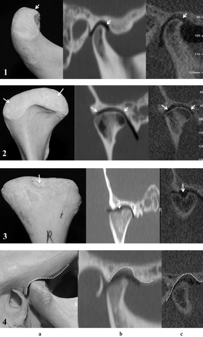
Objective: The aim of this study was to assess the accuracy and reliability of cone beam CT (CBCT) images compared with multidetector CT (MDCT) images for the detection of surface osseous changes in temporomandibular joints (TMJs).
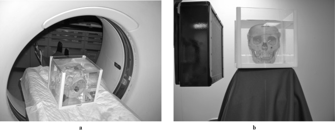
Methods: Naked-eye inspection of 110 sites in 10 TMJs from 5 dry human skulls provided the gold standard. Two radiologists interpreted the images. Sensitivity, specificity and kappa statistics were used for analysis.
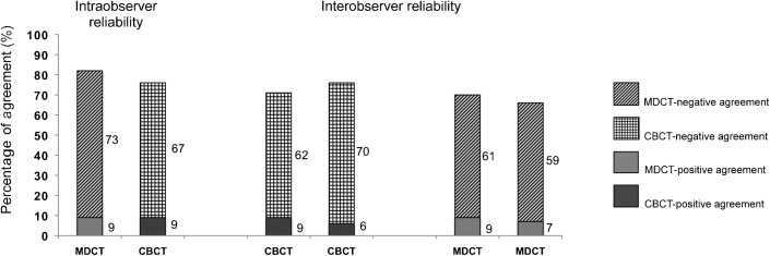
Results: The sensitivities of both modalities were low and comparable whereas the specificities were high and comparable. Intraobserver reliabilities for CBCT (p=0.0005) and for MDCT (p=0.0001) showed significant agreement. Interobserver reliability was higher for CBCT than for MDCT.
Conclusion: CBCT and MDCT accuracy was comparable in detecting surface osseous changes with comparable intraobserver reliabilities. However, since CBCT requires less radiation exposure, it should be encouraged for imaging TMJ with suspected surface osseous changes.
Figures
References
-
- Cohen H, Ross S, Gordon R. Computerized tomography as a guide in the diagnosis of temporomandibular joint disease. J Am Dent Assoc 1985;110:57–60 - PubMed
-
- Lewis EL, Dolwick MF, Abramowicz S, Reeder SL. Contemporary imaging of the temporomandibular joint. Dent Clin North Am 2008;52:875–890, viii - PubMed
-
- Honey OB, Scarfe WC, Hilgers MJ, Klueber K, Silveira AM, Haskell BS, et al. Accuracy of cone-beam computed tomography imaging of the temporomandibular joint: comparisons with panoramic radiology and linear tomography. Am J Orthod Dentofacial Orthop 2007;132:429–438 - PubMed
-
- Cara AC, Gaia BF, Perrella A, Oliveira JX, Lopes PM, Cavalcanti MG. Validity of single- and multislice CT for assessment of mandibular condyle lesions. Dentomaxillofac Radiol 2007;36:24–27 - PubMed
-
- Draenert FG, Coppenrath E, Herzog P, Muller S, Mueller-Lisse UG. Beam hardening artefacts occur in dental implant scans with the NewTom cone beam CT but not with the dental 4-row multidetector CT. Dentomaxillofac Radiol 2007;36:198–203 - PubMed
Publication types
MeSH terms
LinkOut - more resources
Full Text Sources
Medical
