. 2012 Jul;19(7):684-93.
doi: 10.1177/1933719111431000. Epub 2012 Feb 28.

G protein-coupled estrogen receptor (GPER) expression in normal and abnormal endometrium

Affiliations

G protein-coupled estrogen receptor (GPER) expression in normal and abnormal endometrium

Beth J Plante et al. Reprod Sci. 2012 Jul.

Abstract

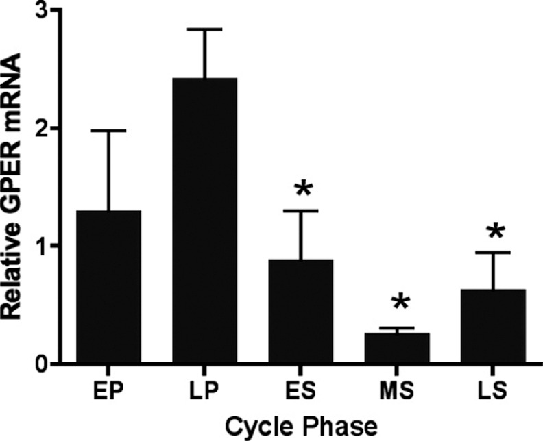
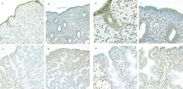
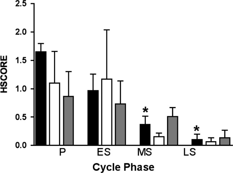
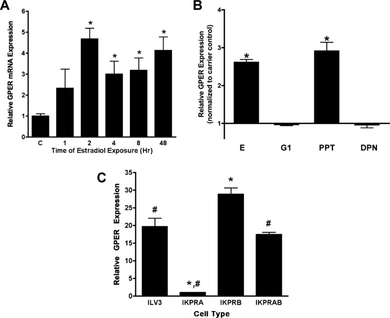
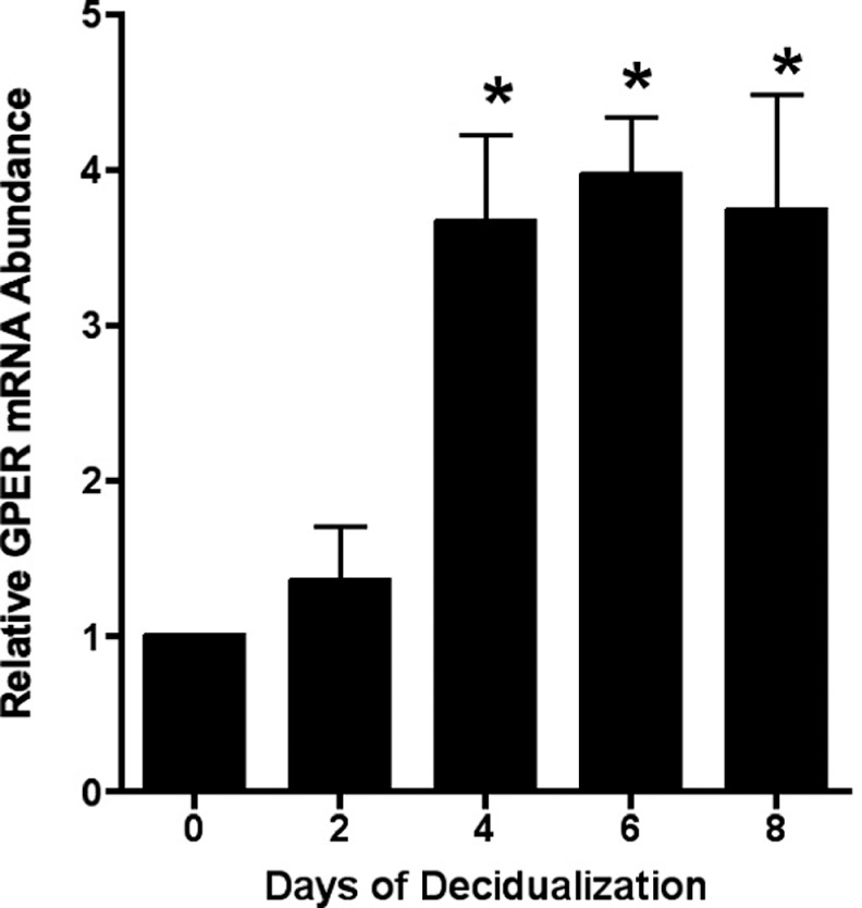
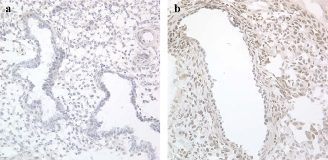
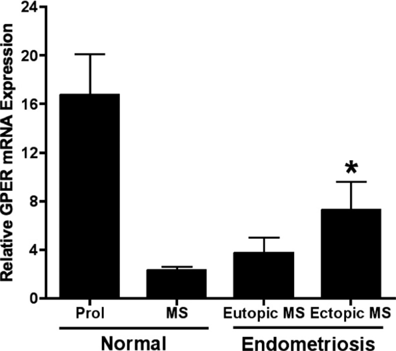
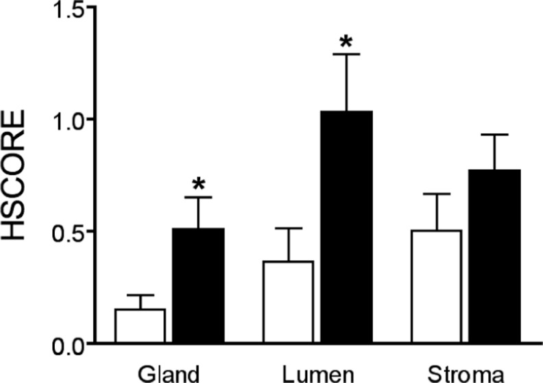
Rapid estrogen effects are mediated by membrane receptors, and evidence suggests a role for both a membrane-associated form of estrogen receptor alpha (ESR1; ERα) and G-protein coupled receptor 30 (GPER; GPR30). Considering estrogen's importance in endometrial physiology and endometriosis pathophysiology, we hypothesized that GPER could be involved in both cyclic changes in endometrial estrogen action and that aberrant expression might be seen in the eutopic endometrium of women with endometriosis. Using real-time reverse transcriptase-polymerase chain reaction (RT-PCR) and immunohistochemical analysis of normal endometrium, endometrial samples demonstrated cycle-regulated expression of GPER, with maximal expression in the proliferative phase. Eutopic and ectopic endometrium from women with endometriosis overexpressed GPER as compared to eutopic endometrium of normal participants. Ishikawa cells, an adenocarcinoma cell line, expressed GPER, with increased expression upon treatment with estrogen or an ESR1 agonist, but not with a GPER-specific agonist. Decreased expression was seen in Ishikawa cells stably transfected with progesterone receptor A. Together, these data suggest that normal endometrial GPER expression is cyclic and regulated by nuclear estrogen and progesterone receptors, while expression is dysregulated in endometriosis.


Conflict of interest statement

Declaration of Conflicting Interests: The author(s) declared no potential conflicts of interest with respect to the research, authorship, and/or publication of this article.

Figures

Figure 1.
Figure 1.
GPER expression in endometrial biopsies from normal women across the menstrual cycle. GPER indicates G protein-coupled estrogen receptor; EP, early proliferative; LP, late proliferative; ES, early secretory; MS, mid-secretory; LS, late secretory; mRNA, messenger RNA. Each bar represents the mean relative mRNA expression at the cycle phase indicated ± standard error of the mean. *P < .05 as compared to late proliferative phase. For EP, LP, ES, MS, and LS, n = 7, 15, 8, 12, and 6, respectively. Data were grouped by cycle phase and analyzed by 1-way analysis of variance using Dunnett Multiple Comparison Test for post hoc analysis.
Figure 2.
Figure 2.
Cyclic changes in immunolocalization of GPER expression in human endometrium. Immunostaining for GPER in human endometrium from proliferative phase (cycle day 10, A and E), early secretory (LH + 3, B and F), mid-secretory (LH+9, C and G), and late secretory (LH + 14, D and H). Panels A-D are taken with a ×20 objective and panels E-H are taken with a ×60 objective and no photos are cropped. GPER indicates G protein-coupled estrogen receptor; LH, luteinizing hormone.
Figure 3.
Figure 3.
GPER HSCORE analysis of immunohistochemical staining in endometrium across the menstrual cycle. Immunohistochemical staining was independently scored in luminal epithelium (black bars), glandular epithelium (white bars), and stroma (gray bars) using the HSCORE method. GPER indicates G protein-coupled estrogen receptor; P, proliferative (n = 3); ES, early secretory (n = 3); MS, mid secretory (n = 16); LS, late secretory (n = 3); HSCORE, histologic scoring. *P < .05 as compared to proliferative phase.
Figure 4.
Figure 4.
Steroid hormone regulation of GPER expression. Panel A, Real-time RT-PCR analysis of GPER mRNA abundance in response to estradiol treatment. Ishikawa cells were treated with 10−8 mol/L E for the indicated times. C, carrier-treated (0.01% ethanol) control. *P < .05 as compared to carrier control. Panel B, Changes in GPER expression in response to E and specific estrogen receptor ligands. Ishikawa cells were treated for 2 hours with 10−8 mol/L E, 10−9 mol/L G1, 10−8 mol/L PPT, 10−9 mol/L DPN, or carrier. Data are expressed as fold change over carrier control. *P < 0.05 as compared to carrier control. Panel C, Effect of PGR on GPER mRNA expression. Ishikawa cells were stably transfected with an empty expression vector (ILV3) or expression vectors for PGR-A (IKPRA), PGR-B (IKPRB), or both PGR-A and PGR-B (IKPRAB), and GPER mRNA was assessed by real-time RT-PCR. *P < .05 as compared to ILV3. # P < .05 as compared to IKPRB. GPER indicates G protein-coupled estrogen receptor; mRNA, messenger RNA; RT-PCR, reverse transcriptase–polymerase chain reaction; E, estradiol; G1, GPER-specific agonist; PPT, propyl pyrazole triol; DPN, diarylpropionitrile; PGR, progesterone receptor.
Figure 5.
Figure 5.
Decidualization increases GPER expression. Isolated human stromal cells were treated with 1 nmol/L estradiol, 100 nmol/L progesterone, and 8-bromo cAMP for the days indicated. Relative quantitation of GPER was performed using real-time RT-PCR. *P < .05 as compared to time 0. GPER indicates G protein-coupled estrogen receptor; RT-PCR, reverse transcriptase–polymerase chain reaction; cAMP, cyclic adenosine monophosphate.
Figure 6.
Figure 6.
Immunolocalization of GPER expression in endometriosis. Immunostaining for GPER from human eutopic (A) and ectopic (B) endometriotic endometrium in the mid-secretory phase. Images are taken with a ×40 objective. GPER indicates G protein-coupled estrogen receptor.
Figure 7.
Figure 7.
Mid-secretory GPER mRNA expression is increased in endometriosis lesions. GPER indicates G protein-coupled estrogen receptor; normal Prol, normal proliferative phase endometrium; normal MS, normal mid-secretory phase endometrium; eutopic MS, eutopic mid-secetory phase endometrium; ectopic MS, ectopic mid-secretory phase endometriosis lesion; mRNA, messenger RNA. Each bar represents the mean relative mRNA expression at the cycle phase indicated ± standard error of the mean. *P < .05 compared to normal mid-secretory phase endometrium. For Normal Prol, Normal MS, Eutopic MS, and Ectopic MS, n = 15, 12, 3, and 3, respectively.
Figure 8.
Figure 8.
Mid-Secretory GPER immunostaining of eutopic endometrium from women with and without endometriosis. Each bar represents the mean HSCORE of the indicated tissue compartment ± standard error of the mean. White bars denote samples from women without endometriosis and black bars denote women with endometriosis. *P < .05 between endometriosis and unaffected samples in the same tissue compartment. For normal glandular tissue, endometriotic glandular tissue, normal luminal tissue, endometriotic luminal tissue, normal stromal tissue, and endometriotic stromal tissue, n = 16, 18, 15, 17, 16, and 18, respectively.

References

    1. Jensen EV, Desombre ER, Hurst DJ, Kawashima T, Jungblut PW. Estrogen-receptor interactions in target tissues. Arch Anat Microsc Morphol Exp. 1967; 56(3): 547–569 - PubMed
    1. Mosselman S, Polman J, Dijkema R. ER beta: identification and characterization of a novel human estrogen receptor. FEBS Lett. 1996; 392(1): 49–53 - PubMed
    1. Jensen EV, Jacobson HI. Basic guides to the mechanism of estrgen action. Recent Prog Horm Res. 1962; 18: 387
    1. Gorski J, Toft D, Shyamala G, Smith D, Notides A. Hormone receptors: studies on the interaction of estrogen with the uterus. Rec Prog Horm Res. 1968; 29: 45. - PubMed
    1. Falkenstein E, Tillmann HC, Christ M, Feuring M, Wehling M. Multiple actions of steroid hormones—a focus on rapid, nongenomic effects. Pharmacol Rev. 2000; 52(4): 513–556 - PubMed

Publication types

MeSH terms