Comparative Study
. 2012 Feb 29;32(9):3176-92.
doi: 10.1523/JNEUROSCI.6439-11.2012.

Inhibition of the NFAT pathway alleviates amyloid β neurotoxicity in a mouse model of Alzheimer's disease

Affiliations
Comparative Study

Inhibition of the NFAT pathway alleviates amyloid β neurotoxicity in a mouse model of Alzheimer's disease

Eloise Hudry et al. J Neurosci. .

Abstract

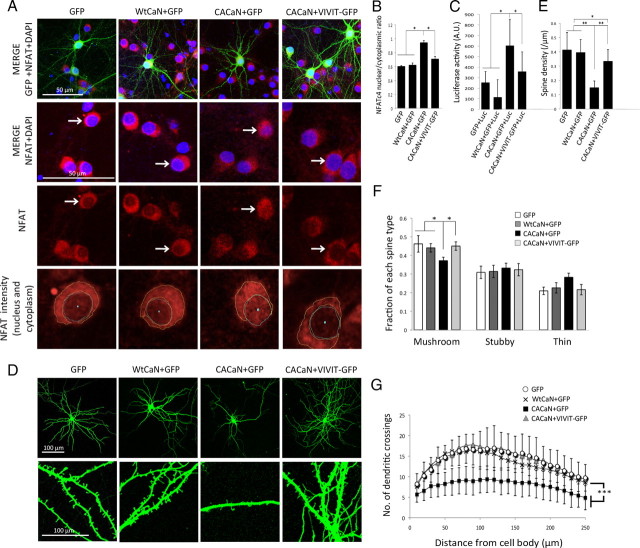
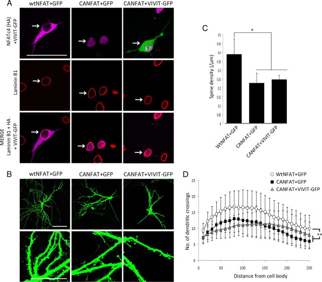
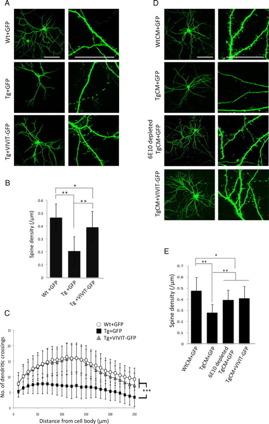
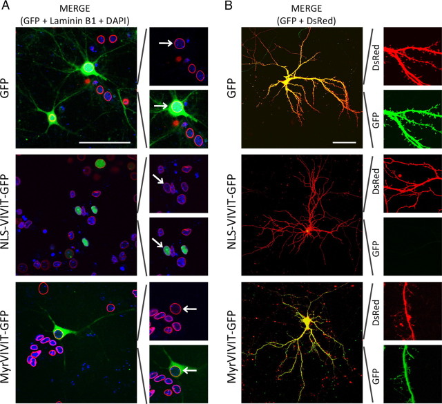
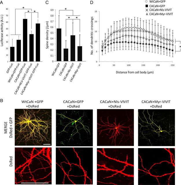
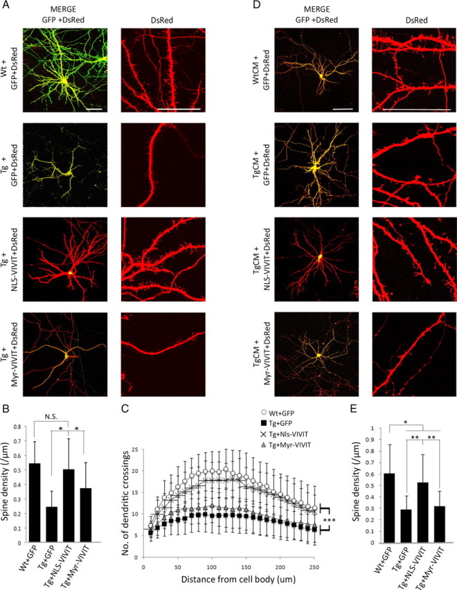
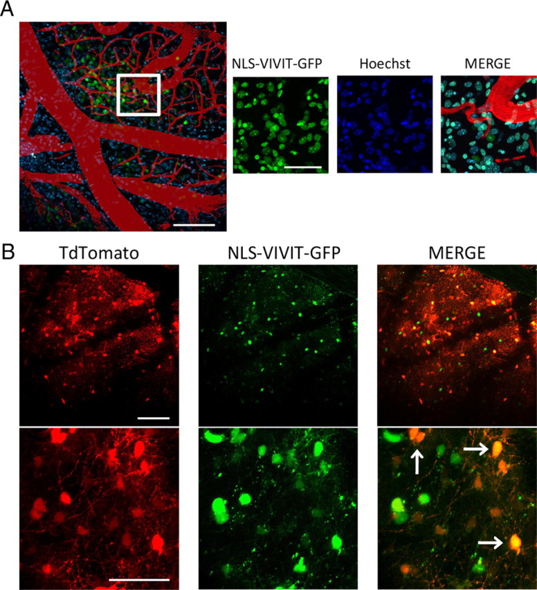
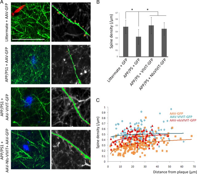
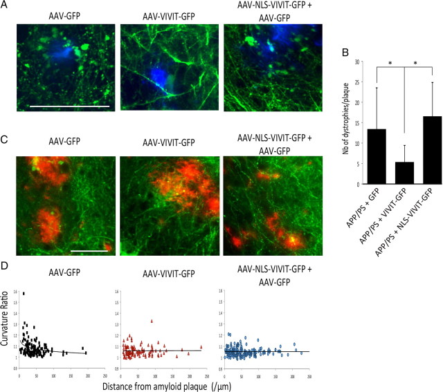
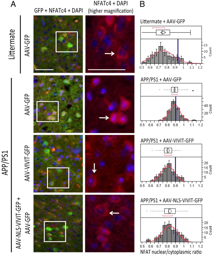
Amyloid β (Aβ) peptides, the main pathological species associated with Alzheimer's disease (AD), disturb intracellular calcium homeostasis, which in turn activates the calcium-dependent phosphatase calcineurin (CaN). CaN activation induced by Aβ leads to pathological morphological changes in neurons, and overexpression of constitutively active calcineurin is sufficient to generate a similar phenotype, even without Aβ. Here, we tested the hypothesis that calcineurin mediates neurodegenerative effects via activation of the nuclear transcription factor of activated T-cells (NFAT). We found that both spine loss and dendritic branching simplification induced by Aβ exposure were mimicked by constitutively active NFAT, and abolished when NFAT activation was blocked using the genetically encoded inhibitor VIVIT. When VIVIT was specifically addressed to the nucleus, identical beneficial effects were observed, thus enforcing the role of NFAT transcriptional activity in Aβ-related neurotoxicity. In vivo, when VIVIT or its nuclear counterpart were overexpressed in a transgenic model of Alzheimer's disease via a gene therapy approach, the spine loss and neuritic abnormalities observed in the vicinity of amyloid plaques were blocked. Overall, these results suggest that NFAT/calcineurin transcriptional cascades contribute to Aβ synaptotoxicity, and may provide a new specific set of pathways for neuroprotective strategies.


Figures

Figure 1.
Figure 1.
VIVIT-GFP overexpression counteracts the effects of constitutively activated calcineurin. A, Representative pictures of GFP, NFATc4 immunostaining, and DAPI (top row) in cortical primary neurons transfected with WtCaN or CACaN, altogether with either GFP or VIVIT-GFP (transfected cells are indicated by an arrow). An increased colocalization of the NFATc4 immunostaining and DAPI is detected in cells overexpressing CACaN and GFP (second row), which is abolished when VIVIT-GFP is cotransfected. The bottom row represents the detection of NFATc4 immunostaining in the nucleus and in the cytoplasm using ImageJ. Scale bar, 50 μm. B, NFATc4 ratios between the nucleus and the cytoplasm are significantly increased when CACaN is overexpressed compared with GFP and WtCaN+GFP. When CACaN is co-transfected with VIVIT-GFP, the nucleus/cytoplasm ratios are significantly decreased and become comparable to neurons transfected with WtCaN+GFP. C, Analysis of NFAT transcriptional activity using the Luciferase reporter system shows that overexpression of CACaN leads to a significant higher Luciferase signal (i.e., higher NFAT transcriptional activity) compared with WtCaN and CACaN+VIVIT. D, E, Representative images and spine density quantification of neurons transfected with GFP, WtCaN/GFP, CACaN/GFP, or CACaN/VIVIT-GFP show that cells co-transfected with CACaN and GFP have a decreased spine density compared with GFP or WtCaN+GFP overexpressing neurons, which is significantly increased by VIVIT-GFP. Scale bar, 100 μm. F, The proportion of each spine type is also affected by CACaN and a significant decrease of the more mature “mushroom” spines is observed compared with GFP and WtCaN/GFP transfected neurons. This effect is greatly abolished when VIVIT-GFP is overexpressed. G, The overall dendritic complexity is also improved when the VIVIT inhibitor was coexpressed with CACaN compared with CACaN/GFP transfected cells. No statistical difference is observed between GFP, WtCaN/GFP, and CACaN/VIVIT-GFP transfected cells. *p < 0.05, **p < 0.001 and ***p < 10−5. (n > 40 cells per condition).
Figure 2.
Figure 2.
Overexpression of a constitutively activated NFATc4 is sufficient to induce abnormal morphological changes in primary neurons. A, Representative images of the nuclear membrane marker laminin B1 and of HA-tagged NFATc4 in cortical neurons that were transduced with WtNFAT/CANFAT (lentiviral vectors). WtNFAT (left) is mainly present in the cytoplasm, outside the nucleus delimited by laminin B1. On the contrary, the constitutively activated form of NFATc4 was concentrated into the nucleus of the cells and fit the laminin B1 staining. The nuclear localization of CANFAT is not affected by the overexpression of VIVIT-GFP (right). Scale bar, 10 μm. B, Images of GFP-filled neurons (top) and dendrites (bottom) that were transduced with either WtNFAT or CANFAT. Scale bar, 100 μm. C, Overexpression of CANFAT is associated with a decreased number of spines per dendritic segment compared with WtNFAT transduced cells. The loss of dendritic spines observed in CANFAT-expressing neurons is not improved by VIVIT-GFP. D, The overall dendritic complexity is also decreased by CANFAT compared with WtNFAT. The lower dendritic complexity induced by CANFAT cannot be rescued by VIVIT-GFP (p > 0.18 when compared with CANFAT/GFP transfected cells). *p < 0.05 and **p < 0.01. (n > 30 cells per condition).
Figure 3.
Figure 3.
The morphological abnormalities observed in Tg neurons and in TgCM-treated neurons are improved by VIVIT-GFP. A, Representative images of Wt and Tg GFP-labeled neurons (left) and dendrites (right) at 18 DIV. Scale bar, 100 μm. B, GFP-transfected Tg neurons present a lower spine density compared with Wt littermate cells, but the number of spines is significantly improved when Tg neurons were previously transfected with VIVIT-GFP. C, In addition, Sholl's plot analyses of Wt and Tg cultures show an overall decreased complexity of the dendritic arborization in Tg neurons overexpressing GFP compared with Wt neurons. The simplification of the dendritic arbor is fully corrected when Tg neurons were transfected by VIVIT-GFP and it reaches the level of complexity observed in Wt neurons (p = NS compared with Wt neurons transfected with GFP). D, Representative images of wild-type cortical neurons (left) and dendrites (right) expressing GFP or VIVIT-GFP after treatment for 24 h with WtCM, TgCM, or depleted TgCM. Scale bar, 100 μm. E, Dendritic spine quantification demonstrates that a short exposure to oligomeric Aβ leads to a significant decrease of their density, whereas spines are unaffected when WtCM was applied. However, the spine density is maintained when TgCM-treated neurons overexpress VIVIT-GFP or when Aβ peptides were previously immunodepleted from the medium using the 6E10 antibody. *p < 0.01, **p < 0.0001 and ***p < 10−10. (n > 40 cells per condition).
Figure 4.
Figure 4.
NLS-VIVIT-GFP localization is restricted to the nucleus, whereas Myr-VIVIT-GFP is excluded from this compartment. A, Representative images of primary neurons that were transfected with GFP, NLS-VIVIT-GFP, or Myr-VIVIT-GFP and then stained for laminin B1 and DAPI (left). On the right, higher magnification images show that GFP alone can be detected in the nuclear and cytoplasmic compartments, whereas NLS-VIVIT-GFP is surrounded by laminin B1 and colocalizes with DAPI, suggesting that it is restricted to the nucleus. Inversely, Myr-VIVIT-GFP is excluded from the nuclear compartment. Scale bar, 50 μm. B, Both GFP and Myr-VIVIT-GFP can be detected in the dendritic shaft and in spines, as suggesting by the colocalization with DsRed. NLS-VIVIT-GFP is, however, absent in the neuronal processes. Scale bar, 100 μm.
Figure 5.
Figure 5.
The restricted localization of VIVIT into the nucleus potently inhibits the morphological abnormalities induced by an overactivated form of CaN. A, NFAT transcriptional activity assessed using the pNFAT-luciferase reporter system shows that overexpression of NLS-VIVIT-GFP, altogether with CACaN, is associated with a decreased luciferase activity. No effect is observed when Myr-VIVIT-GFP is overexpressed, suggesting that this inhibitor does not affect NFAT transcriptional activity. B, Representative images of cortical primary neurons (top) and dendrites (bottom) that were co-transfected with GFP, NLS-VIVIT-GFP, and Myr-VIVIT-GFP altogether with CACaN and WtCaN. To be able to observe the neurites and quantify the dendritic spines in all the conditions, cells were co-transfected with a DsRed fluorescent reporter. Scale bar, 100 μm. C, The decreased spine density associated with the overexpression of a constitutively activated CaN (CACaN) can be improved by co-transfection with a nuclear-targeted VIVIT (NLS-VIVIT-GFP), whereas no beneficial effects is observed with a membrane-bound VIVIT. D, Overexpression of NLS-VIVIT also leads to a more complex dendritic arborization of CACaN transfected neurons, whereas overexpression of Myr-VIVIT cannot rescue the effects of CACaN. *p < 0.05. (n > 30 cells per condition).
Figure 6.
Figure 6.
Aberrant neuronal morphology observed in Tg neurons and in TgCM-treated cells are prevented by NLS-VIVIT-GFP but not by Myr-VIVIT-GFP. A, Representative images of DsRed-filled Wt or Tg neurons (left) and dendrites (right) overexpressing GFP or the different VIVIT constructs show that Tg cells transfected by NLS-VIVIT-GFP present an increased spine density compared with GFP transfected Tg cells. Scale bar, 100 μm. B, Bar graph summarizing the averaged spine density of each group confirms that only the nuclear targeted peptide is able to improve the morphological abnormalities observed in Tg neurons, but this is not the case when Myr-VIVIT-GFP is overexpressed. C, Sholl's plot analysis demonstrates that NLS-VIVIT-GFP also increases the dendritic complexity compared with GFP-transfected Tg neurons and reaches the level of complexity observed in Wt neurons. D, Images representative of DsRed filled neurons (left) or dendritic segments (right) after co-transfection by GFP, NLS-VIVIT-GFP, or Myr-VIVIT-GFP and 24 h treatment with WtCM and TgCM. Scale bar, 100 μm. E, A decreased spine density was induced by TgCM, which could be inhibited by NLS-VIVIT-GFP but not by Myr-VIVIT-GFP. *p < 0.05, **p < 0.001 and ***p < 10−5. (n > 40 cells per condition).
Figure 7.
Figure 7.
In vivo, NLS-VIVIT-GFP is targeted to the nucleus and coinjection of both AAV-NLS-VIVIT and AAV-Tdtomato efficiently transduces the same cells. A, Representative in vivo images of a mouse injected with AAV-NLS-VIVIT-GFP (left) confirm that the localization of NLS-VIVIT is restricted to the nucleus and colocalizes with Hoechst applied topically (right). Scale bars: 100 μm on the left and 50 μm on the right. B, Coinjection of AAV-NLS-VIVIT-GFP and AAV-Tdtomato with an appropriate ratio (3:1) is an efficient approach to be able to detect spines in vivo and to ascertain the fact that the observed Tomato-transduced cells also express NLS-VIVIT (arrows indicate GFP/Tdtomato double positive cells). Scale bars: 100 μm on the top and 50 μm on the bottom.
Figure 8.
Figure 8.
Spine loss detected in the vicinity of amyloid deposits is significantly improved by VIVIT-GFP and NLS-VIVIT-GFP. A, Left, z-projections of two-photon images representative of GFP-filled neurites in littermate mice injected with AAV-GFP or surrounding amyloid deposits in APP/PS1 mice injected with AAV-GFP, AAV-VIVIT-GFP, and AAV-NLS-VIVIT-GFP+AAV-GFP. Right, Pictures represent an example of dendritic spine detection using the Neuronstudio software. Brown, red, and yellow circles respectively indicate mushroom, stubby, and thin spines. Scale bar, 50 μm. B, Bar graph summarizing the averaged spine density of each group confirms that APP/PS1 mice injected with AAV-VIVIT-GFP and AAV-NLS-VIVIT-GFP have a higher spine density than AAV-GFP injected Tg animals within 100 μm surrounding amyloid deposits. Spine densities of either VIVIT-GFP or NLS-VIVIT-GFP treated groups are not significantly different from GFP injected littermates. C, Scatter plot of spine density data collected from GFP-injected APP/PS1 mice show a positive correlation between spine density and distance from amyloid deposit, but this local effect of amyloid plaque is partially and even completely abolished when animals were treated with NLS-VIVIT-GFP or VIVIT-GFP, respectively. *p < 0.05 between AAV-GFP-treated APP/PS1 mice and either AAV-VIVIT-GFP or AAV-NLS-VIVIT-GFP injected animals. (n = 6 animals and n < 200 dendrites analyzed per condition).
Figure 9.
Figure 9.
Overexpression of VIVIT-GFP and NLS-VIVIT-GFP partially improve the neuritic abnormalities in vivo. Representative pictures (A) and quantifications (B) of dystrophies surrounding amyloid deposits show that fewer neuritic dystrophies are present when AAV-VIVIT-GFP is injected, but no improvement can be observed when NLS-VIVIT-GFP is overexpressed. Scale bar, 50 μm. (n < 35 amyloid plaques and n < 250 dystrophies analyzed per group). C, Representative images of GFP and amyloid deposits immunostaining in APP/PS1-injected mice. Scale bar, 100 μm. D, In APP/PS1 mice injected with AAV-GFP, the increased curvature ratio is inversely correlated with the distance of each neurite to the closest amyloid deposit (p < 0.0001, slope =-0.0006). There is, however, no such correlation in APP/PS1 mice treated with either VIVIT-GFP (p = 0.953, slope =-5.12 × 10⁻⁶) or NLS-VIVIT-GFP (p = 0.812, slope = 0.00003). *p < 0.05. (n = 6 animals per group).
Figure 10.
Figure 10.
Overexpression of VIVIT-GFP and NLS-VIVIT-GFP prevent NFATc4 accumulation in the nucleus of transduced neurons. A, Representative images of GFP immunostaining and endogenous NFATc4 in injected animals allows detection of endogenous NFATc4 in transduced neurons. Scale bar, 50 μm; for higher magnification images, on the right, 20 μm. Higher magnification images of NFATc4 and DAPI (third column) show that an increased recruitment of NFATc4 is detected in AAV-GFP-injected APP/PS1 mice. B, The ratios of NFATc4 immunofluorescence intensity between the nucleus and the cytoplasm follow a normal distribution in all three groups tested. However, there is a shift toward higher values in AAV-GFP-injected APP/PS1 mice compared with AAV-VIVIT and AAV-NLS-VIVIT treated animals. On the graph, the blue line represents the mean of NFATc4 nuclear/cytoplasmic ratio in AAV-GFP injected APP/PS1 mice. The nuclear/cytoplasmic ratio is significantly increased in AAV-GFP treated transgenic mice compared with AAV-VIVIT-GFP and AAV-NLS-VIVIT-GFP (p < 0.05). (n < 4 animals per group and 3 slices per animal were analyzed).

Comment in

References

    1. Abdul HM, Sama MA, Furman JL, Mathis DM, Beckett TL, Weidner AM, Patel ES, Baig I, Murphy MP, LeVine H, 3rd, Kraner SD, Norris CM. Cognitive decline in Alzheimer's disease is associated with selective changes in calcineurin/NFAT signaling. J Neurosci. 2009;29:12957–12969. - PMC - PubMed
    1. Alvarez S, Blanco A, Fresno M, Muñoz-Fernández MÁ. TNF-alpha contributes to caspase-3 independent apoptosis in neuroblastoma cells: role of NFAT. PLoS One. 2011;6:e16100. - PMC - PubMed
    1. Amberg GC, Rossow CF, Navedo MF, Santana LF. NFATc3 regulates Kv2.1 expression in arterial smooth muscle. J Biol Chem. 2004;279:47326–47334. - PubMed
    1. Aramburu J, Yaffe MB, López-Rodríguez C, Cantley LC, Hogan PG, Rao A. Affinity-driven peptide selection of an NFAT inhibitor more selective than cyclosporin A. Science. 1999;285:2129–2133. - PubMed
    1. Bacskai BJ, Klunk WE, Mathis CA, Hyman BT. Imaging amyloid-beta deposits in vivo. J Cereb Blood Flow Metab. 2002;22:1035–1041. - PubMed

Publication types