. 2012 May 1;72(9):2440-53.
doi: 10.1158/0008-5472.CAN-11-4038. Epub 2012 Feb 29.

Suppression of the epithelial-mesenchymal transition by Grainyhead-like-2

Affiliations

Suppression of the epithelial-mesenchymal transition by Grainyhead-like-2

Benjamin Cieply et al. Cancer Res. .

Abstract

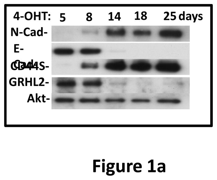
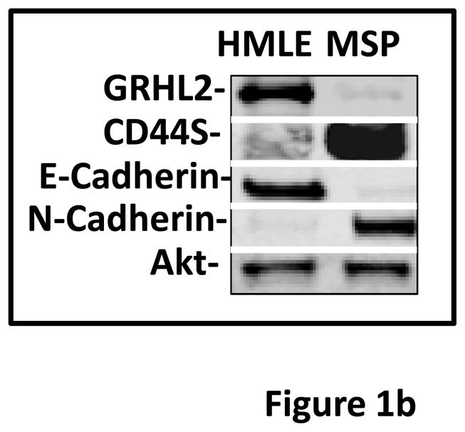
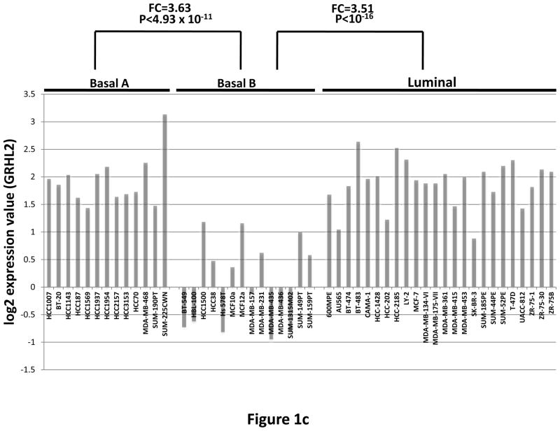
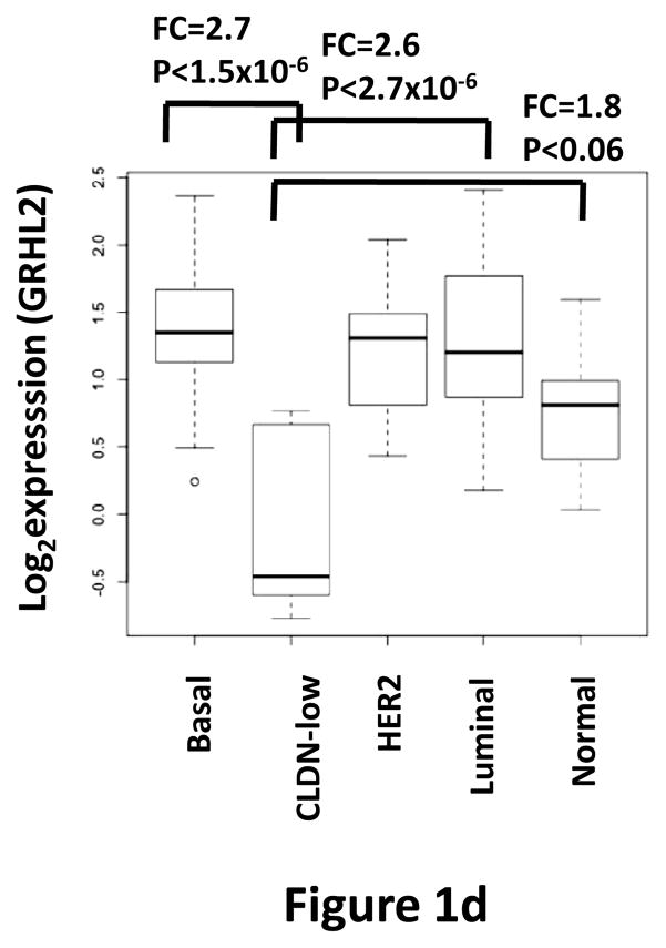
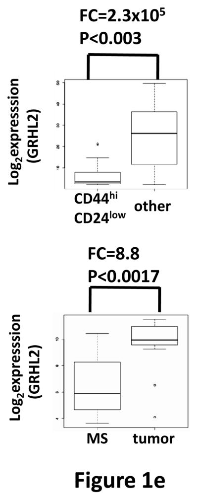
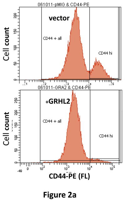
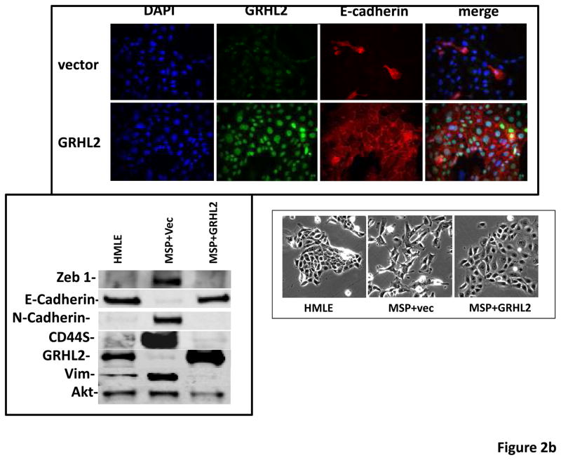
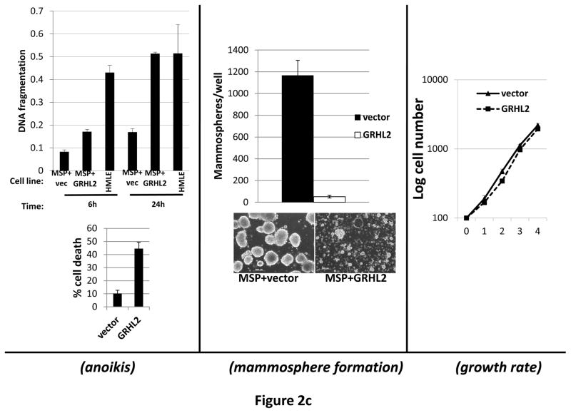
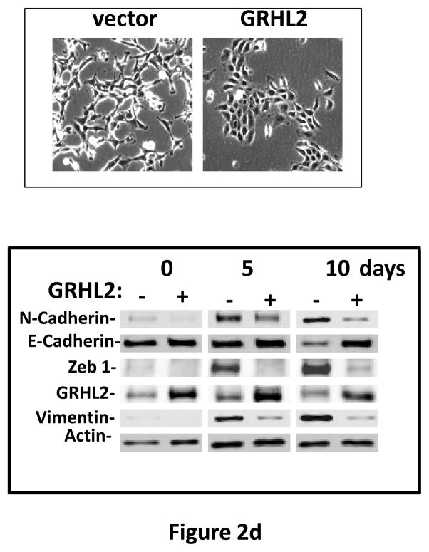
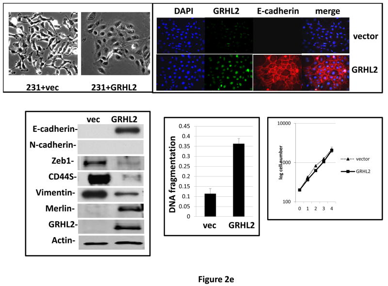
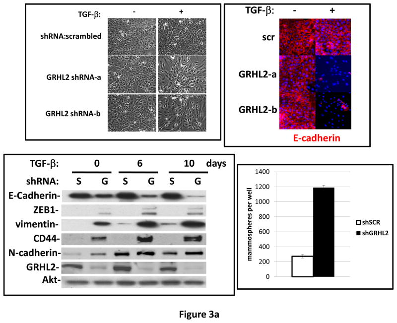
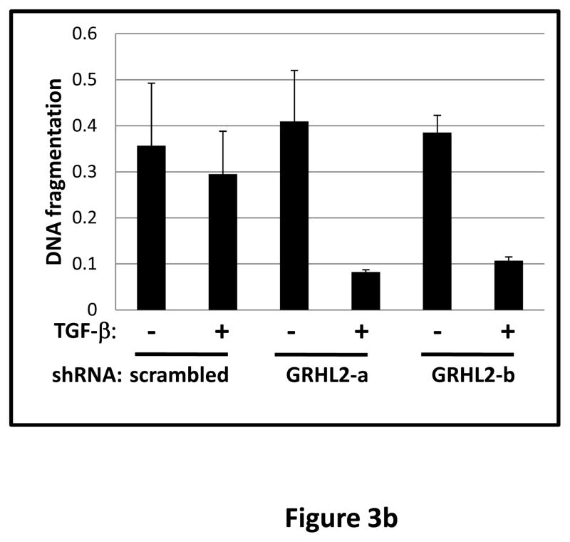
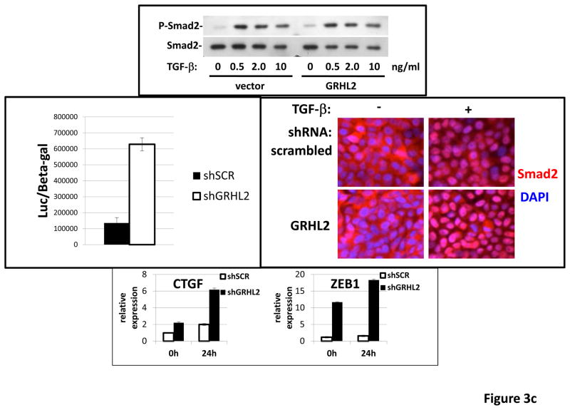
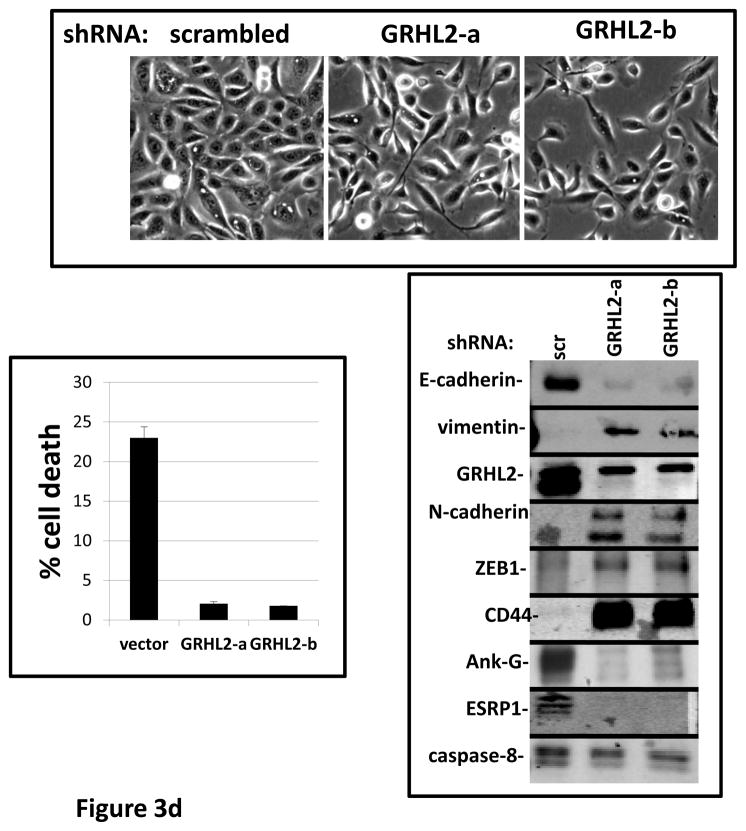
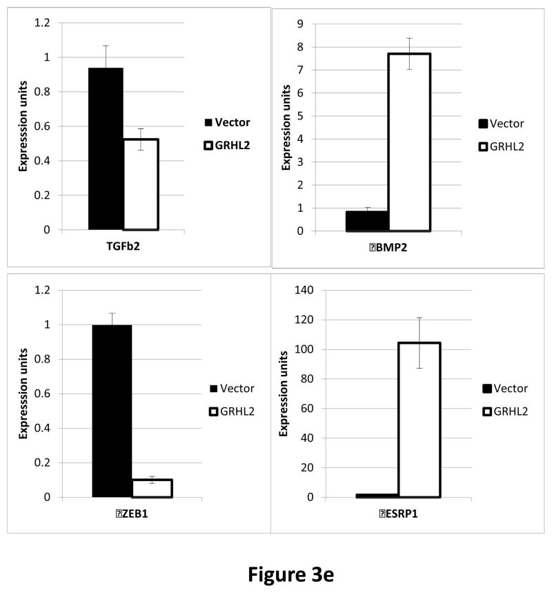
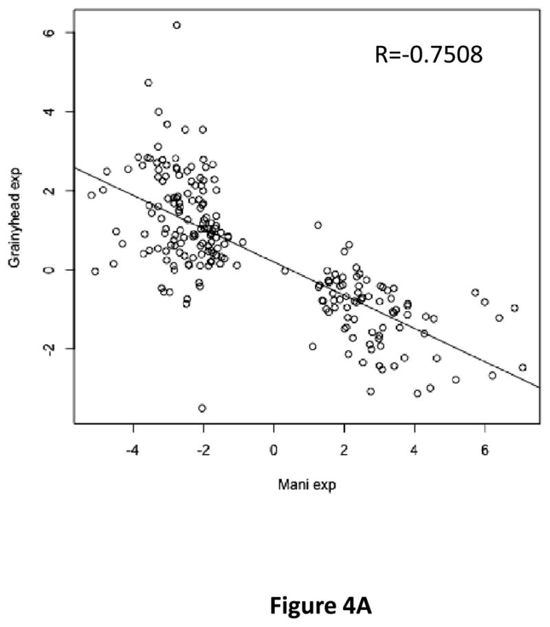
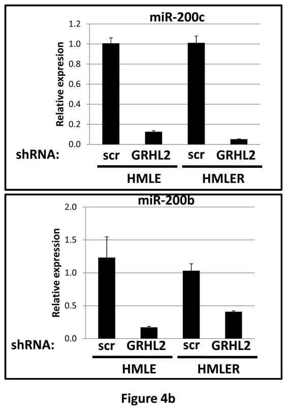
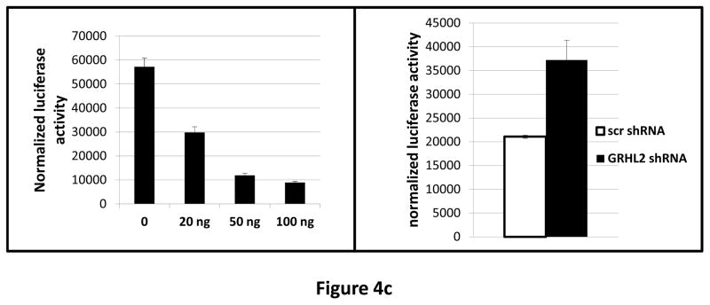
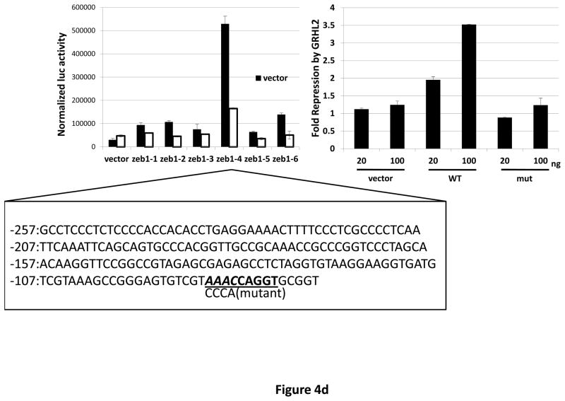
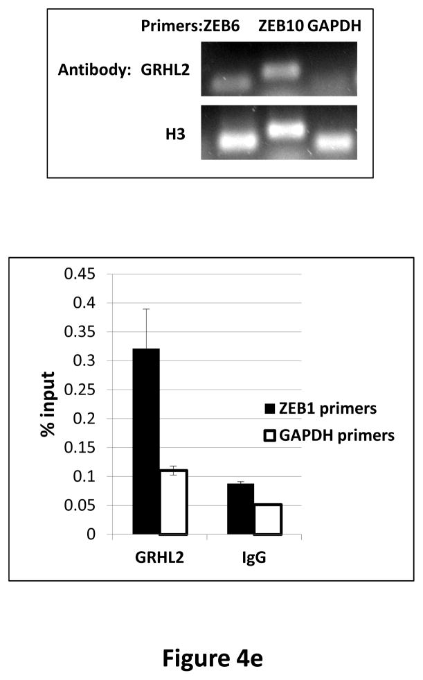
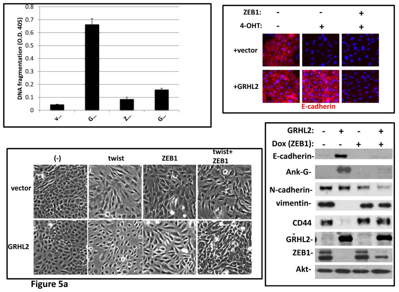
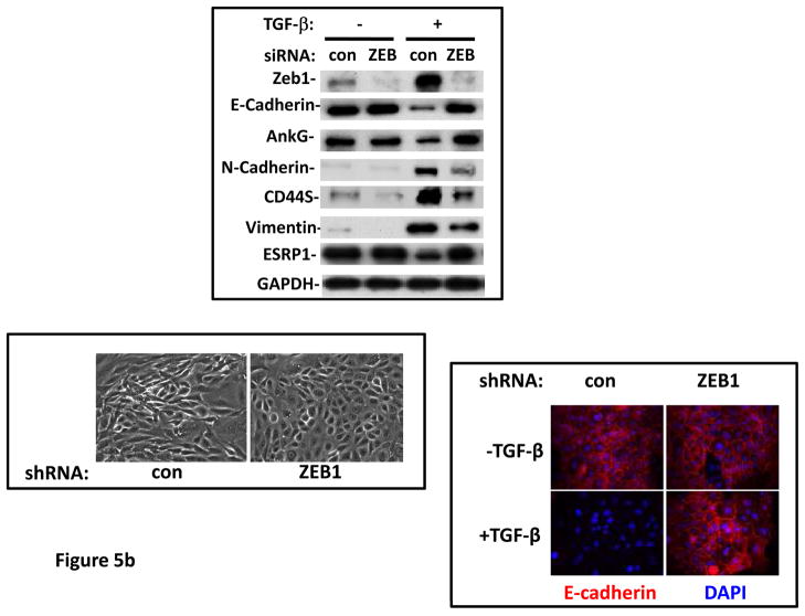
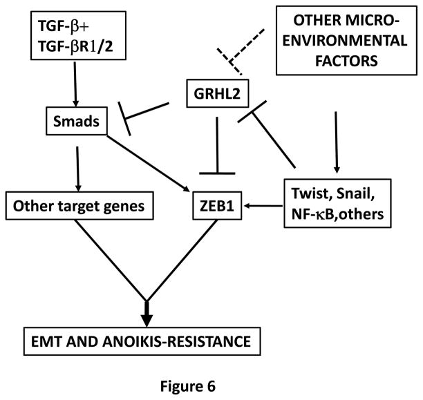
Grainyhead genes are involved in wound healing and developmental neural tube closure. In light of the high degree of similarity between the epithelial-mesenchymal transitions (EMT) occurring in wound-healing processes and the cancer stem cell-like compartment of tumors, including TGF-β dependence, we investigated the role of the Grainyhead gene, Grainyhead-like-2 (GRHL2) in oncogenic EMT. GRHL2 was downregulated specifically in the claudin-low subclass breast tumors and in basal-B subclass breast cancer cell lines. GRHL2 suppressed TGF-β-induced, Twist-induced or spontaneous EMT, enhanced anoikis sensitivity, and suppressed mammosphere generation in mammary epithelial cells. These effects were mediated in part by suppression of ZEB1 expression via direct repression of the ZEB1 promoter. GRHL2 also inhibited Smad-mediated transcription and it upregulated mir-200b/c as well as the TGF-β receptor antagonist, BMP2. Finally, ectopic expression of GRHL2 in MDA-MB-231 breast cancer cells triggered an MET and restored sensitivity to anoikis. Taken together, our findings define a major role for GRHL2 in the suppression of oncogenic EMT in breast cancer cells.


Figures

Figure 1
Figure 1. GRHL2 is down-regulated in cells and tumors that have undergone EMT
(a) Twist down-regulates GRHL2. HMLE cells with tamoxifen-inducible twist (twist-ER) were induced with 4-hydroxytamoxifen for the indicated periods of time and analyzed for epithelial, mesenchymal and tumor intiating cell (CD44) marker genes. (b) GRHL2 is down-regulated in the Mesenchymal Sub-Population (MSP) cells relative to parental HMLE cells. (c) GRHL2 is down-regulated specifically in “basal B” subclass of breast cancer cell lines characterized by Neve, et al. (42). (d) GRHL2 is down-regulated specifically in the “claudin-low” subclass of breast cancer cell lines characterized by Hennessy, et al. (3). (e) GRHL2 is down-regulated specifically in the tumor initiating/mesenchymal cell subpopulation characterized by Creighton, et al (25).
Figure 2
Figure 2. GRHL2 suppresses EMT
(a) Ectopic expression of GRHL2 suppresses the CD44 expression in the spontaneously occurring mesenchymal subpopulation of HMLE cells (“MSP”). HMLE+Twist-ER cells (without 4-OHT) infected with GRHL2 or empty retroviral vector (pMIG) were analyzed for CD44 expression by FACS. (b) GRHL2 reverts MSP cells to an epithelial phenotype. The CD44high mesenchymal sub-population of HMLE, obtained by flow-sorting of HMLE cells, were infected with empty vector or GRHL2 and then (top panel) stained for the indicated proteins by immunofluorescence or (lower panel) probed for epithelial and mesenchymal marker genes by western blotting. (c) GRHL2 expression in MSP cells restores anoikis-sensitivity (left panels: top graph represents a DNA fragmentation ELISA assay, lower graph represents a trypan blue-permeability assay) and reduces mammosphere (middle panels), without affecting growth rate (right panel). (d) GRHL2 suppresses Twist-induced EMT. HMLE+twist-ER expressing either empty vector or GRHL2 were treated with 4-OHT (to activate the twist transgene) for 7 days, and (top panel) photographed to record morphology. (lower panel): Time course of changes in epithelial and mesenchymal marker genes after induction of the twist gene with 4-OHT, in cells with or without ectopic GRHL2 expression; (e) GRHL2 suppresses EMT and anoikis-resistance in MDA-MB-231LN cells. MDA-MB-231LN expressing either empty vector or GRHL2 were analyzed for morphology (phase contrast microscopy, top left), E-cadherin expression and localization (immunofluorescence, top right), expression of epithelial and mesenchymal markers (western blotting, bottom left), anoikis-sensitivity (DNA fragmentation ELISA (middle) and adherent growth rate (lower right).
Figure 3
Figure 3. GRHL2 suppresses TGF-β-induced EMT
(a) GRHL2 knockdown permits HMLE cells to undergo TGF-β-induced EMT. HMLE+twist-ER cells (no 4-OHT) with two distinct GRHL2 shRNAs or a scrambled control shRNA were exposed to TGF-β for two weeks prior to analysis for cell morphology (top left), E-cadherin expression/localization (immunofluorescence, top right) or mammosphere generation (graph, lower right). Epithelial and mesenchymal markers were also analyzed at the indicated times of TGF-β treatment (western blot, lower left). (b) GRHL2 suppresses the ability of TGF-β to confer anoikis-resistance. The cell lines described above were assayed for anoikis by DNA fragmentation after two weeks incubation with or without TGF-β. (c) GRHL2 inhibits Smad-mediated transcription. (left panel): Effect of stable GRHL2 knockdown on activity of a Smad-responsive reporter construct (3TP-lux) was determined by luciferase assays of transiently transfected HMLE+twist-ER cells (no 4-OHT) expressing either scrambled or GRHL2a shRNA that were treated with TGF-β for sixteen hours prior to lysis; values are luciferase activity normalized to an internal co-transfected β-galactosidase control. (lower panel): Effect of stable GRHL2 knockdown on induction of TGF-β/Smad target genes, CTGF and ZEB1, by exogenous TGF-β, determined by qRT-PCR. (top panel): No effect of GRHL2 on phosphorylation of Smad2/3. MSP cells expressing empty vector or GRHL2 were treated with the indicated concentrations of TGF-β for 24h and analyzed for total and phospho-smad2/3. (right panel): No effect of GRHL2 on nuclear translocation of Smad2. The GRHL2 knockdown or control cells described in part a were treated with TGF-β for six hours and analyzed for Smad2 localization by immunofluorescence. (d) GRHL2 knockdown induces EMT in HMLE cells with Ras (HMLER). HMLER expressing two distinct GRHL2 shRNAs or scrambled shRNA control were imaged using phase contrast microscopy (top), immunoblotted for epithelial and mesenchymal markers (right) or assayed for anoikis (trypan blue exclusion, left panel). (e) GRHL2 up-regulates BMP2. RT-PCR was performed on RNAs from HMLE+Twist-ER cell lines with or without constitutive GRHL2 expression (induced with 4-OHT), using the indicated primer sets.
Figure 4
Figure 4. GRHL2 represses the ZEB1 gene
(a) HMLE+Twist-ER expressing either empty vector or GRHL2 were induced with 4-OHT for 17 days; four days following removal, RNAs were isolated and compared by microarray profiling. Gene changes due to GRHL2 were compared to those due to Twist in the same cell line; a regression plot of this comparison is shown. (b) GRHL2 up-regulates mir-200b/c. RNAs from HMLE or HMLER cells expressing GRHL2 or control shRNAs were compared for the indicated mir-200 transcripts by RT-PCR. (c) GRHL2 represses the ZEB1 promoter. (left panel): MSP cells were co-transfected with a ZEB1 promoter-luciferase reporter construct, with the indicated input amounts of GRHL2 expression vector or equal amounts of empty vector. Values represent the relative luciferase activity normalized to an internal β-galactosidase control; (right panel): HMLE+Twist-ER cells (no 4-OHT) with stable GRHL2 knockdown or control (scrambled) knockdown were assayed for luciferase activity after transient transfection of the ZEB1 promoter. (d) Identification of a ZEB1 promoter fragment that is GRHL2-sensitive, and contains a GRHL2 consensus binding site. (left panel): Fragments (~200bp) spanning the 1 kb ZEB1 promoter were assayed for transcriptional activity in the presence or absence of co-transfected GRHL2. The fragment 1–4 sequence and predicted GRHL2 binding site are shown. (right panel): Fragment 4 containing wild-type or mutant versions of the predicted GRHL2 binding site were assayed for repression by co-transfected GRHL2. (e) GRHL2 protein interacts with the ZEB1 promoter. Chromatin from HMLE+Twist-ER+GRHL2 (upper panel) or MDA-MB-231+GRHL2 (lower panel) was immunoprecipitated with GRHL2, histone H3 or non-immune antibody and analyzed by gel-based PCR or qPCR, using the indicated CHIP primers.
Figure 5
Figure 5. Suppression of ZEB1 is important for the suppression of EMT by GRHL2
(a) ZEB1 restores EMT capability and anoikis-resistance in cells that express GRHL2 constitutively. HMLE+twist-ER with or without constitutive GRHL2 expression and with empty vector or ectopic ZEB1 expression vector were treated with 4OHT and then analyzed for anoikis (DNA fragmentation ELISA, top left); E-cadherin expression/localization (immunofluorescence, top right), cell morphology (phase contrast, lower left) and expression of epithelial and mesenchymal markers (western blot, lower right). (b) ZEB1 knockdown prevents TGF-β-induced EMT in GRHL2 knockdown cells. HMLE+twistER (without 4OHT) expressing shGRHL2a were transfected with either non-targeting siRNA or ZEB1 siRNA prior to incubation with or without TGF-β Cells were analyzed for epithelial and mesenchymal markers (western blot, top panel), morphology (phase contrast, lower left panel), or E-cadherin expression/localization (immunoflourescence, lower right).
Figure 6
Figure 6. Summary of the proposed model
EMT is induced by the combination of TGF-β with other micro-environmental factors, and requires the activation of ZEB1 plus other target genes. Microenvironmental factors (Wnt, NF-kb agonists?) up-regulate Twist and Snail genes, which down-regulate GRHL2. This down-regulation alleviates the repression of the ZEB1 promoter, permitting TGF-β (partly, through Twist and Snail themselves) to activate ZEB1 expression. The down-regulation of GRHL2 also enhances Smad-mediated transactivation of TGF-β target genes, which, together with ZEB1, induce EMT and anoikis-resistance.

References

    1. Polyak K, Weinberg RA. Transitions between epithelial and mesenchymal states: acquisition of malignant and stem cell traits. Nat Rev Cancer. 2009;9:265–73. - PubMed
    1. Thiery JP, Acloque H, Huang RY, Nieto MA. Epithelial-mesenchymal transitions in development and disease. Cell. 2009;139:871–90. - PubMed
    1. Hennessy BT, Gonzalez-Angulo AM, Stemke-Hale K, et al. Characterization of a naturally occurring breast cancer subset enriched in epithelial-to-mesenchymal transition and stem cell characteristics. Cancer Res. 2009;69:4116–24. - PMC - PubMed
    1. Massague J. TGFbeta in Cancer. Cell. 2008;134:215–30. - PMC - PubMed
    1. Taylor MA, Parvani JG, Schiemann WP. The pathophysiology of epithelial-mesenchymal transition induced by transforming growth factor-beta in normal and malignant mammary epithelial cells. J Mammary Gland Biol Neoplasia. 15:169–90. - PMC - PubMed

Publication types

MeSH terms